CHAPTER 2. Anatomy of the ZOR
■ Development of eyes
■ Ochnitsa
■ Very apple
zovnishnya obolonka
Middle shell
Internal shell (sitkivka)
Vmist full-time apple
bloodstending
інnervatsіya
Glyadachev Shlyakh
■ Additional equipment of the eye
okorukhovikh m'yazi
births
conjunctiva
sl_zni organi
OCHI ROSVITOK
The rudiment of an eye appears in the 22-day embryo in the viglyadi bet of small invaginations (full-time furrows) in the forebrain. Gradually intussusception grows and forms virility - vernacular belly. The distal part of the intrauterine mikhur is pressed into the ear of the n'yat intrauterine development, establishing the intrauterine cells. The name of the wall of a full-time cell gives an ear to the copy of the copy of the copy, and the internal - to the copy of the copy of the copy.
At the stage of full-time mihurіv in the adjoining dіlyankas of the ectoderma, there is a cultivation - krishtalikov plakoіdi. It is possible to see the molding of the crystal bulbs and drawing them into the empty cells of the inner cells, while the front and rear cameras of the eye are formed. The ectoderm above the full cell also gives an ear to the ear of the horn.
In mesenchymal cells, bezposeredno navkolishny intramural cells, a sudin hemispheres develop and a sudin shell is formed.
Neuroglial elements give an ear of myoneural tissue and sphinkter and dilator of the brain. The nazovn_ from the vesicular sheath from the mesenchymal develops the nasal fibrous unformed tissue of the sclera. Ahead of it, the projection of the foresight and the transition to the connecting-fabric part of the horn.
In the end of another month of the ectoderm, the development of the sleazy ones. Okorukhovy m'yazy rozvayutsya from myotomes, presenting a transversely dark m'yazovoy tissue of the somatic type. The workers will fix the shape of the folds. The stench grows smartly nasustrich one to one and grows between oneself. Behind them, there is a space as if to be seen by the prismatic epithelium, a conjunctival bear. On the 7th month of the intrauterine development, the conjunctival beetle starts to develop. Along the edge of the capital, there are vії, greasy and vidozminenі sweats.
Features of the eyes of children
In new-born women, the apple is very large, but rather short. The residual size of the eyes will rise up to 7-8 years. Novonarodzheniy can be very large and large flat, lower in older adults, rogivka. When populated, the shape of the crystal is spherical; stretching the living life in the growth and becoming more flat, it is poured into the statements of new fibers. Newborns in the stroma of the ridge have little to no pigment, but not much. Blakytny color of the eyes nadaє evoked posterior pigment epithelium. If the pigment is repaired in the parenchyma of the ridge, you will see the new color.
ochnytsi
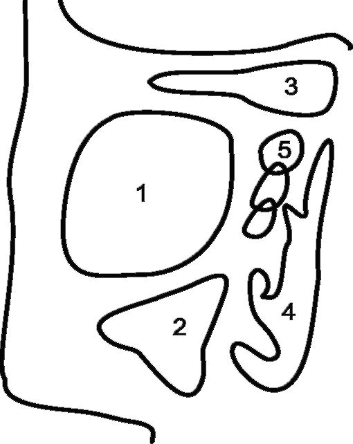
Orbita(Orbita), for the sake of it, is a guy who has been set at the viglyad of a loss in the anterior part of the skull, which is nagadu chotirigrannu pyramid, the top of which is straight back and a spike to the middle (Fig. 2.1). Cleansing inside, top, bottom and bottom walls.
The inner wall of the orbit is represented by an even thinner plate, as it is empty from the middle of a thin cluster. With a single fee for the sinus, you can easily go into the orbit and to the child of the poop who has gone through his emphysema. The top-inner
Small. 2.1.Budova orbit: 1 - superior orbital fissure; 2 - small crust of the main brush; 3 - channel of the zorotic nerve; 4 - rear openings; 5 - orbital plate of the grating brush; 6 - front lacrimal ridge; 7 - flaky brush and back flaky comb; 8 - the pit of the teardrop; 9 - nasal brush; 10 - frontal outgrowth; 11 - lower ochnoyamkovy edge (upper slit); 12 - bottom slot; 13 - low-eyed boron; 14. infraorbital opening; 15 - lower orbital cleft; 16 - wicker basket; 17 - round opening; 18 - great crust of the main brush; 19 - lobova brush; 20 - upper intra-pitched edge
The small orbit of the orbit is between the frontal sinus, and the lower part of the orbit is from the maxillary sinus (Fig. 2.2). Tse of enhancing the expansion of ignition and swelling processes from the paranasal sinuses into the orbit.
The lower part of the orbital is often done with blunt injuries. A direct blow to the full-time apple of the wicker, pushing the grip in orbit, and the lower side of it “fail”, flushing when there is a cyst defect in the edge of the eyelet.
Small. 2.2.Orbit and pre-sinus: 1 - orbit; 2 - maxillary sinus; 3 - forehead sinus; 4 - nasal walk; 5 - sinus cavity
Tarzo-orbital fascia and advances on this very apple serve as a front wall, which will surround the empty orbit. The tarzoorbital fascia is attached to the edges of the orbit and cartilage of the capital and is tightly tied with a tenon capsule, which curls the apple from the lamb to the healthy nerve. In front of the Tenon's capsule is zednana with conjunctiva and episcleritis, and behind the Kremlin, the apple is seen from the orbital cell. Tenon's capsule is suitable for all okorukh muses.
Basically, instead of orbit - fat cells and okorukhovyh oysters, the very apple itself borrows a part of the orbit's obsyag. All the illumination, roztasovani in advance of the tarzoorbital fascia, to lie in the posture ochnitsa (zokrema, sloppy bear).
Ochnitsa tie with empty skull hello to help you decorate openings.
The superior orbital fissure is closed by the orbital opening with the middle cranial fossa. Such nerves pass through it: okoruchiy (III pair of cranial nerves), blocky (IV pair of cranial nerves), ochnoyamkovy (first pair of cranial nerves) and double (VI pair of cranial nerves). The upper internal vein, the main vessel behind the roof of the internal apple and orbital, also passes through the upper internal section.
Pathology in the area of the upper orbital cleft can lead to the development of the syndrome of "upper orbital cleft": ptosis, general malfunction of the intraocular apple (ophthalmoplegia), mediasis, parallax amoodocity, impaired sensibility viniknennya eksophthalmu.
Vіdnya orbit through the upper internal slit to pass into the empty space of the skull and fall into the cavernous sinus. Anastomoses with the veins of an individual, perch for everything through the angular vein, and also the visibility of the venous valves, take a wide spread of infection from the upper part of the exposure into the orbit and distal to the emptying of the skull with the development of thrombotic sinus cavity.
The lower orbital fissure is from the lower orbital gap from the cryopidnebine and the lateral-lower slit pits. The lower orbital gap is closed with a full tissue, in the yak weaved smooth fibers. When the nice innervations of the meat are destroyed, the vinikє enophthalm (the oche-
foot apple). So, with urazhenny fibers, go to the top of the sympathetic vuznogo vuznogo vuznogo pit, Horner's syndrome develops: partial ptosis, myosis and enophthalmos. The canal of the zorovaya nerve of the rosette at the apex of the ochnitsa in the small krill of the main cyst. Through the whole canal to enter the empty space of the skull, the healthy nerve and enter the orbit of the internal artery - the blood supply of the eye and the additional apparatus was mostly dull.
VERY APPLE
It’s very hard to store an apple from three shells (first, middle and inner) and together (nasty body, kryshtalik, as well as the native vology of the anterior and posterior chambers of the eye, Fig. 2.3).
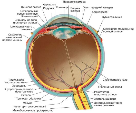
Small. 2.3.Scheme of the budovi intramural apple (sagital spriz).
zovnishnya obolonka
Zovnishnya, abo fibrous, shell of an eye (Tunica fibrosa) presented by rogivka (Cornea)і sclera (Sclera).
cornea - the foresight of the out-of-court part of the outer shell of the eye. The function of the flashing is the carrying out and breaking of light changes, as well as the clearing of the in-person apple from the unwelcoming, inviting injections. The diameter of the corrugation should be set to the middle 11.0 mm, the thickness - from 0.5 mm (in the center) to 1.0 mm, the breaking point is close to 43.0 diopters. In the norm, the horny sheath is open, smooth, blinking, spherical and highly sensitive to tissue. The influx of unwelcoming inviting factors on the horn of the wicker is the reflexive squeezing of the table, the insensibility of the internal apple (horn reflex).
The cornea is stored in 5 balls: anterior epithelium, Bowman's membrane, stroma, Descemet's membrane and posterior epithelium.
front Large-ball flat necrophilia of the epithelium of the viconus function and in case of injury, the regeneration is increased by holding out.
bowman membrane- the basal membrane of the anterior epithelium. Vona st_yka to mechanical injections.
Stroma(Parenchyma) horns warehouseє up to 90% її goods. It is stored from thin thin plates, between bright and flexible cells and a great number of sensitive nerve ends.
"Descemet's membrane є the basal membrane of the posterior epithelium. Vaughn to serve as an overhead bar on the way of widening infections.
back drink to be stored in one ball of hexagonal form. Vin pereshkozhaє a reliable lead from the anterior chamber to the stroma of the horn, but not regenerated.
Harchuvannya of the corneous membrane is seen for the shell of the pericorneal fissure of the sudin, in the anterior chamber of the eye і sleep. The transparency of the horn is enveloped in a one-sided structure, in the presence of the court and is strictly signified by the wickedness of water.
limb- mіsce transition of the cornea into the sclera. Tse napivprozorium rim, width is close to 0.75-1.0 mm. The company has a limba for the shuffling of the Schlemm canal. Limb serve as a garnish guard in the inventory of pathological processes in the brain and sclera, as well as in the case of surgical intervention.
Bilkova- the opacity of a part of the outer shell of the eye, where there is a large colorectal shell (bilkovy shell). The width of the syaga is 1 mm, and the naythonsha part of the sclera is welded into the path of the external nerve. The functions of the sclera are written and form-creating. Bilkova for her bud is similar to the parenchyma of the corneal membrane, protea, as seen from her, filled with water (through the visibility of the epithelial cover) and opacity. Numerous nerves and judges pass through the sclera.
Middle shell
The middle (sudinna) shell of the eye, or the uveal tract (Tunica vasculosa), to be stored in three parts: rajduzhki (Iris), ciliary body (Corpus ciliare)і goodіdeі (Choroidea).
Raiduzhna Obolonka serve as an automatic diaphragm of the eyes. Tovshchina ryadzhki only 0.2-0.4 mm, naymenha - in the middle of the transition to the ciliary body, so you can get rid of the rails in case of injuries (іdodіaliz). The raid is stored from the half-tissue stroma, the sudin, the epithelium, which curls the raid in front and two balls of the epithelium behind the back, preventing the opacity. The stroma of the rye shell is to avenge the lack of blood chromatophores, a bit of melanin in certain types of eyes. The ryaduzhny obolontsi have a remarkably small number of sensitive nerve ends, so the fiery ridge of the ridge is superfluous with a painful syndrome.
zinitsya- a round hole in the center of the rail. The workers of the change of their diameter regulate the number of light changes that fall on the screen. The value of the interval changes from two smooth muzzles of the ridge shell - sphincter and dilator. M'yazov fibers of the sphincter roztashovani ring-like and perceive parasympathetic innervations from the okorulovy nerve. The radial fibers of the dilator are innervated from the upper shyny nice vnod.
tsiliarne tilo- a part of the sheath of the eye, yak at the viglyad, the ring passes through the root of the ridge and the choroid. The cordon between the ciliary body and the choroid passes along the tooth line. Cylillarne tilo viroblyaє internal іdina and take part in the act of accommodation. In the area of cylindrical growths, there is a good deed. At the ciliary school, the establishment of an internal family is being introduced. ciliary
it is stored from decilkokh bundles of rizno-straightened fibers, which is attached to the sclera. Fast and forward, the stench will loosen the tightness of the price links, as you go from the cylindrical spine to the capsule of the crystal. When the cylinder body is ignited, the process of accommodation will be damaged. The innervation of the ciliary body is sensitive (I head of the tripartite nerve), parasympathetic and sympathetic fibers. In the ciliary type, there are significantly more sensitive nerve fibers, lower in the ridge, and in this case, when the painful swelling syndrome is inflamed. horioideya- the posterior part of the uveal tract, adjoined to the ciliary body of the toothed line. The good news is to be stored from decilkoh balls of the Sudin. The ball of wide choriocapillaries adheres to the sieve and appears from it by Bruch's thin membrane. The name of the ball of the middle Sudins (above all arterioles), behind which there is the ball of the Bigger Sudins (venules). Between the sclera and choroid є the suprachoroidal space, in which transit pass the judgment and nerves. In the choroid, as in the upper portions of the uveal tract, pigment cells develop. The good news is that you will not be able to eat the new spheres of the brain (neuroepithelium). There is blood in the choroid of hope, as a result of the diagnosis of metastatic swells and the occurrence of diseases of children in infectious diseases. Goodness is not rejecting sensitive energy, so good things are painlessly opposed.
Internal shell (sitkivka)
The inner shell of the eye is represented by a retina - high-coded differentiated nerve tissue, designated for the sprinkling of light podraznikiv. From the disc of the healthy nerve to the dentate line, the optically effective part of the sieve is formed, as it is stored from the neurosensory and pigment ball. In front of the toothed line, rosette at 6-7 mm from the limba, it is reduced to the food, which covers the ciliary body and the iris. The part of the sittings does not take part in the act.
The sieve is grafted from the choroid only along the toothed line in front and near the disk of the healthy nerve and along the edge of the zhovtoy beach behind. The thickness of the sieve should be close to 0.4 mm, and in the area of the tooth line and in the zhovty plyami - only 0.07-0.08 mm. grabbing the sits
Welcome to the show of good ideas and central art of the art. Sitkivka, as a good idea, is not a little energy.
The functional center of the sitk is the zhovta plyama (macula), which is a courtless dilyanka of a rounded form, the number of changes in lutein and zeaxanthin. Naybilsh svitlochutlivaya part of the zhovtoy plyami - the central fossa, abo foveoli (Fig. 2.4).
Budovi sitkivka scheme
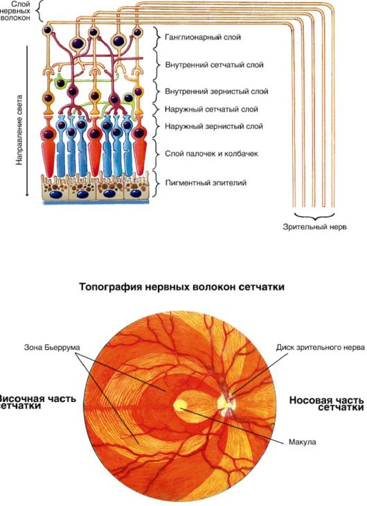
Small. 2.4.Scheme of budovi sitskivka. Topography of nerve fibers
In the group of 3 first neurons of the healthy analyzer: photoreceptors (first neuron) - sticks and cones, bipolar cells (another neuron) and ganglion cells (third neuron). The sticks and cones are the receptor part of the healthy analyzer and are located in the last balls of the sieve, without the middle in the middle of the dietary intake. sticks, roztasvani on the periphery, leading to the peripheral vision - the field of vision and light perception. cones, The main mass of zooseredzhena in the region is zhovtoyi beaches, the central zir (state property zoru) and kolorovidchuttya are provided.
Visoka razdіlnіst zhovtoї beaches amumbled with such features.
Do not go through the picture here and do not overshoot the change of light on the photoreceptor.
At the central pit, only cones grow, all the balls of the stems are brought to the periphery, allowing the exchange of light to be consumed directly on the cones.
Particularly, the spіvvіdnoshennya neurons of the sieve: in the central pit, one bipolar clitin per seizure cone, and on the cutaneous bipolar clitin - its own ganglionic clitin. Thus, there is a “direct” connection between photoreceptors and healthy centers.
On the periphery of the sieve, navpaki, one bipolar cline is brought to the stick, and one ganglion cline is added to the bipolar clump. The summation of the subtlety of the peripheral part of the sieve is highly sensitive to the smallest amount of light.
The axons of ganglionic cells converge, establishing a healthy nerve. The disc of the healthy nerve leads to the movement of nerve fibers from the inside of the apple and does not take revenge on light-sensitive elements.
Vmist full-time apple
Instead of a full-time apple - sclopodibne tilo (Corpus vitreum), kryshtalik (Lens), as well as the watery vologue of the front and back chambers of the eye (Humor aquosus).
Sklovidne tilo for a vagoya y obyag to become approximately 2/3 of the full-time apple. Tse the hole of the courtless draglist illumination, memorizing the space between the sieve, the cylindrical body, the fibers of the zinc ring and the crystal. It is confined to them by a thin near-cord membrane, in the middle of which there is a skeleton of
thin fibrils і gel-shaped tongue. It is less likely to be stored by 99% from water, in which case there is a small amount of alcohol, hyaluronic acid and electrolytes. It is not easy to finish it is tied to the ciliary body, the capsule of the crystal, as well as to the suture close to the tooth line and in the area of the green nerve disc. In the wake of the sound of the capsule of the crystal is weak.
kryshtalik(Linza) - a hole, without a court-like elasticity, which has the shape of a double-convex linse with a thickness of 4-5 mm and a diameter of 9-10 mm. The river of kristalik with a firm consistency is packed in a thin capsule. The functions of the kristalik are carrying out and breaking light changes, as well as participating in accommodation. The force of breaking the crystal to become close to 18-19 diopters, and at the maximum pressure of accommodation - up to 30-33 diopters.
Krishtalik grows beside the middle behind the iris and suspensions on the fibers of the zinc link, which are inserted into the capsule of the crystal at the yogi. Equator razdіlya kristalik capsule on the front and back. Krym tsyogo, krishtalik has an anterior and a posterior pole.
From the front capsule of the crystal, subcapsular feeds are formed, which produces fiber by stretching the living life. At the same time, the Krishtalik grows more flat and strong, consuming their elasticity. The building is gradually absorbed before the accommodation, so as the speech of a Kristalik cannot change its shape. Krishtalik may be 65% more stored in water, and instead of a small one, 35% - more, less in any kind of fabric of our body. Linz also has a smaller number of mineral speech, ascorbic acid and glutathione.
internal ridin to be produced in the cylindrical room, to store the front and back chambers of the eye.
The anterior chamber of the eye is a space between rogivkoyu, rayduzhkoyu and kryshtalik.
The back chamber of the eye is a vuzka shchіlina mіzh іzhka і kryshtalik with a zinn ring.
watery vologa Take care of the fate in the harchuvanny of the unjudged middle-class eyes, and the exchange in the meaning of the world is the value of the internal vice. The main path to the inner ridini is the cut of the anterior chamber of the eye, the statement is made by the root of the ridge and the horny sheath. Through the system of trabeculae and the ball of the inner tube of the inner line, go into the Schlemm canal (venous sinus), the ips are seen at the vein of the sclera.
bloodstending
All arterial roofs come close to Yabluko in full-time arteries (A. Ophthalmica)- internal sleepy arteries. Full-time artery from such gilki, how to go to full-time apple:
Central artery of the sieve, which will prevent the blood flow of the internal balls of the sieve;
The posterior short ciliary artery (number 6-12), dichotomously reddened in the choroid and superimposed on blood;
The back of the ciliary artery (2), which passes into the suprachoroidal space to the ciliary body;
The anterior ciliary artery (4-6) is seen from the muscular artery.
The posterior and anterior ciliary arteries, anastomosed with each other, create a great arterial colo ridge membrane. From the direction of the radial, the judges come in, to form the arterial colo near the zone of the malium. For the rakhunok of the posterior and anterior ciliary arteries, the ridge and the ciliary body will become sick with blood, the pericorneal hemispheres of the sudins will become established, take care of the fate of the crustal cornea. Alone the bloodsteps, you change your mind for an hour-long firing of the ridge shell and the cylindrical body, at that hour, when you are good at it, you will be isolated.
The blood from the intramural apple is seen through the vortic (vortex) veins, anterior ciliary veins and the central veins of the stench. Vorticus veins take shelter from the uveal tract and fill the very apple, obliquely piercing the sclera near the eye. Anterior ciliary vein and central vein
інnervatsіya
I am very sensitive, sympathetic and parasympathetic.
sensitive energy to take care of the orbital nerve (the I stub of the tripartite nerve), which in the empty orbit is 3 stubs:
Loose and supra-well nerves, which do not seem to be able to lead to an innervating apple;
The nasal nerve is formed by 3-4 of the ciliary nerve, which can pass without the middle in the ovum of the apple, and also take care of the fate of the formed ciliary nerve.
tsilіarniy vuzolseams in 7-10 mm from the posterior pole of the intramuscular apple and lagging to the external nerve. The ciliarny vuzol has three corintsia:
Sensitive (from the nasal nerve);
Parasympathetic (the fibers are blown out at once with the okorukhovy nerve);
Nice (from fibers of cute gossip). From the ciliary university to go to the full-time apple 4-6 short
ciliary nerves. Before them, adhere to the nice fibers, go to the dilator of the zinitsi (the stench does not go into the ciliar vuzol). In such a rank, the short ciliary nerves of the brain, in the mind of the young ciliary nerves, carry only sensitive fibers.
Short and long ciliary nerves go up to the posterior pole of the eye, pierce the sclera and go into the suprachoroidal space to the ciliary body. Here the stench comes from sensitive spirits to the railroad, horny and ciliary body. One of the innervations of signs of the eye is accumulated during the development of a single symptom complex - horny syndrome (sloughing, pituitary and blepharospasm). From the most common ciliary nerves, it is also possible to see the sympathetic and parasympathetic nerves to the muses of the brain and the ciliary body.
Glyadachev Shlyakh
Glyadachev Shlyakhthey are stored from healthy nerves, healthy crossing, healthy paths, as well as adult and bright green centers (Fig. 2.5).
Healthy nerve (n. Opticus, II pair of cranial nerves) are formed from the axons of ganglionic neurons of the sieve. On the day of the day, the disc of the healthy nerve is only 1.5 mm in diameter and the body is physically thin - the slap of the plyama. If the apple is very close, the healthy nerve will remove the cerebral sheaths and enter the empty skull through the channel of the healthy nerve.
zorove crossover (Chiasma) to form when the inner halves of the healthy nerves melt. At the same time, there are healthy paths, which place the fibers from the new ones of the same eyes and fibers, so that they go out from the inner half of the anti-aging eyes.
Pіdkіrkovі zorovі centri roztashovani in the most common areas, the axon of ganglionic cells will end. fiber
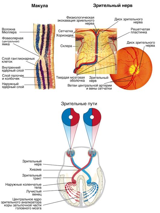
Small. 2.5.Scheme of budovy zorovikh nobles, zorovy nerve and sits
the central neuron through the posterior stigto-internal capsule and the Graziole bundle to go to the measles clit in the cavernous part in the spur furrow (cortical viddil zorovogo analyzer).
ADDITIONAL APPARATUS OCHI
Before the additional apparatus of the eye, it is necessary to carry out okorukhovi muzyazi, loose organisms (Fig. 2.6), as well as symptoms and conjunctiva.
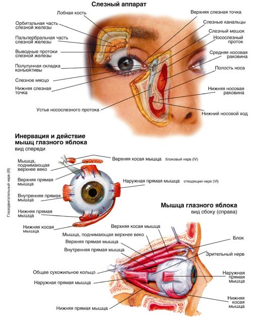
Small. 2.6.Budova of sl_znih organs and meat apparatus of an intramural apple
okorukhovikh m'yazi
Okorukhovyh m'yazi will get rid of the crumbling apple. Oh shist: chotiri straight and two oblique.
Straight joints (upper, lower, outer and inner) are repaired from the tendon ring of the Cinna, the rosette bele of the apex of the orbit near the healthy nerve, and is attached to the sclera 5-8 mm from the limbus.
The upper oblique hole should be repaired from the side of the internal fossa from the top and up to the middle from the opening, from forward, to be thrown over the block, straight from the back and from the bottom, to the sclera in the upper 16 mm quadrant.
The lower oblique muzzle should be repaired from the medial pattern of the orbit behind the inferior orbital fissure and attached to the sclera in the inferior-outer quadrant 16 mm from the limb.
The name of the straight line, where the eyes are brought in, is innervated by the nerve (VI pair of cranial nerves). The upper oblique m'yaz, the tendon which spreads over the block, is the trochlear nerve (IV pair of cranial nerves). The upper, inner and lower straight, as well as the lower braid of the muzzle are innervated by the okoruchial nerve (III pair of cranial nerves). The blood posture of okorukhovyh mu'yazіv zd_ysnyutsya mu'azovyh gilkami full-time artery.
Diya okorukhovyh mu'yazi: the inner and the last of the straight mu'yazi turn the apple very horizontally in the direction of the same name. Upper and lower straight - in a vertical straight line to the sides of the same name and to the middle. The upper and lower braids of the tongue turn the eyes to the side, the opposite with the name of the tongue (so that the upper one goes to the bottom, and the lower one - burns down), і named. Coordinating six pairs of okorukhovyh mu'yaziv will prevent binocular zir. In the destruction of the functions of the languages (for example, with paresis or parallel to one of them), the double function of one eye will be affected.
births
births- crumbling shkirno-m'yazov folds, scho zakrivayut very much the apple call. The stench to seize the eye from the ear, the excess of light, and the moment of additional help, in the same way, curl it up with the slice of water
corneous membrane and conjunctiva Worms are stored in two spheres: anterior - nodular-myasial and posterior - mucous-cartilaginous.
cartilage- solid nasal fibrous plates, which form a table, to be located between themselves at the inner and outer root of the tendon adhesions. On the big edge of the capital, there are two ribs - front and back. The space between them is called intermarginal, the width of its component is approximately 2 mm. At the end of the space, the ducts of the meibomian zalosis, which have grown in the cartilage, are visible. At the forefront of the capital are the roots of Zeiss' oily hairs and species of Moll's salmon. At the medial kut full-time slits on the posterior ribs of the table there are lingering points.
shkira vikeven thinner, the puff of the crumbs is fluffy and does not take revenge on the fatty tissue. It is easier to explain the diagnosis of capital growth in case of early muscular ailments and systemic pathology (heart-vascular, nirkov, etc.). In case of fractures of the ophthalmic cysts, the walls of the paranasal sinuses can be fixed, so that the child can use the development of his emphysema.
M'azi stolittya.At the fabrics of the stol_ttya, a circular thread of the eye is roasted. With the speed of the turn, the turnouts begin to swell. M'yaz is the innervus of the facial nerve, with the development of lagophthalmos (non-micanisation of intramural spines) and vivoritis of the lower vertex. At the top of the top dressing, you will also have the same type of dressing as the top dressing. Vona repair at the top of the orbit and in three portions fit into the skin of the capital, its cartilage and conjunctiva. The middle part of the ointment is innervated by the fibers of the upper part of the pretty stovbur. To that, with a damaged sympathetic nervosa, partial ptosis (one of the manifestations of Horner's syndrome). Reshta of a part of the mucus, which pіdnіmaє the upper band, іnnervatsіyu from the okorulovy nerve.
bloodstending stolittya zd_ysnyuєtsya with full-time arteries. The vascularisation is even more good, and the reason for this is that they have a high reparative health. The lymphatic view from the upper condition goes to the previous lymphatic universities, and from the lower one - to the secondary. Sensitive energy of the capital is provided with I and II throats of the tripartite nerve.
conjunctiva
conjunctivaIt is a thin membrane, embedded in a large ball of epithelium. We see the conjunctiva of the full-time apple (curving the front surface behind the vignette of the horn), the conjunctiva of the transition folds and the conjunctiva of the capital (whistling the back surface).
Subsequent tissue in the area of transitional folds reveals the number of adenoid elements and lymphoid cells, which make follicles. There is no evidence of conjunctiva in the norm of follicles. At the conjunctiva of the upper transitional folds, additional tears of the Krause folds and ducts of the main lacrimal folds appear. Bagatosharovy cylindrical epithelium of the conjunctiva is seen as mucin, which in the storehouse of lacrimal pill covers the horny membrane and conjunctiva.
Blood posture of the conjunctiva from the system of the anterior ciliary arteries and arterial vessels of the capital. Lymphatic education from con'n'unks - motives to stay up to the earliest and most advanced lymphatic universities. Sensitive to the innervation of the conjunctiva, it will take care of I and II throats of the tripartite nerve.
sl_zni organi
To the lacrimal organs to carry the tear-producing apparatus and the lacrimal way.
Tear-producing apparatus (Fig. 2.7). The main driftwood grows in the drip hole in the upper-final form of the orbit. At the upper conjunctival star, there are ducts (about 10), the main lacrimal gulfs and the diseaseless other additional laxative valleys of the Krause and Wolfring. Have zvychaynyh minds for zvolozhennya full-time apple to complete the functios of additional lazy vines. Sleep (main) start to function with uncomfortable cold influxes and actions of emotional camps, which can be manifested as laxity. Bloodstaining from a tearful artery, from a tearful artery, from blood to being seen in a vein ochnytsi. Lymphatic judges from lazy zalozi go to the previous lymphatic universities. The innervation of the lazy nerve is connected to the first nerve of the tripartite nerve, as well as by the pretty nerve fibers from the upper, sympathetic, sympathetic university.
Lacrimal Way. Going into the conjunctival star of the tears of the birthplace of the zavdyak Then it’s easy to get away from the vuz space between the lower whim and the full apple - a tearful stream, the stars go to the teary lake in the medial groove of the eye. At the bottom of the lake, the upper and lower lagging points are covered with holes, they are roasted on the medical part of the countryside. From the lazy points of the tear, come near the upper and lower lazy tubules, which fall into the lazy little shock. A sloppy little trick rots into the pose of emptying the orbit at the inner kut in the yamts. Distant tears come up into the nasolacrimal duct, which ascends into the lower nasal path.
Slosa. When it comes to storage in the main water, and also to take revenge on the bottles (including immunoglobulins), lysozyme, glucose, K +, Na + and Cl - and the other components. The pH is normal at mid-range 7.35. Slyza takes care of the fate in the establishment of the slippery pudding, as it oberіga the surface of the intramural apple from the visihannya and іnfіkuvannya. Slip the mat thickness of 7-10 microns and be stored in three balls. Superficial - a ball of lipids to the secret of meibomian zalosis. Wine is confident in the vaping of a slavish birth. The middle ball is itself a slink of the middle. The internal ball is to take revenge on mucin, so that the cells of the cell-like cells of the conjunctivi are rocked.
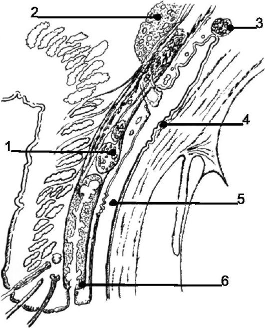
Small. 2.7.Tear-producing apparatus: 1 - Wolfring's zalozi; 2 - slinky hair; 3 - zaliza Krause; 4 - zalozi Manza; 5 - crypt Henle; 6 - vivіdny potіk meibomievy zalozi