Normal anatomy to the organ of zoru people │ Part 1
1-12-2012, 11:50
Healthy analyzer of people The organism is referred to the sensory systems in the anatomical and functional manner, it is stored from decile interconnections, albeit for the main characteristics of structural units (Fig. 3.1):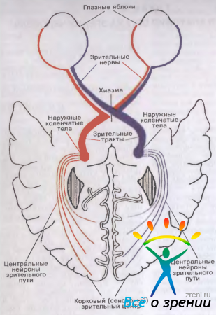
Small. 3.1. Budov's zorovogo analyzer of people (diagram).
- two apples, grown at the frontal area in the right and in the skin, with an optical system, allowing focusing on the sieve (wholly of the receptor part of the analyzer), the image of all the areas of their call systems
- systems of life and safety of the structures of the analyzer (blood circulation, innervation, innervating of the inner life, regulation of the fluid and hemodynamics).
Ochne apple (bulbus oculi)
human eye, Approximately 2/3 of the seams when empty, the shape is not correct. In healthy newborns of the same size, with a path of growth, equal (in the middle) along the sagittal axis 17 mm, transverse 17 mm and vertical 16.5 mm. In older people with dim refractive eyes, the indicators become 24.4; 23.8 and 23.5 mm inconspicuous. Masa of a full-time apple of a new-born is located in the boundaries of up to 3 g, a growth of people - up to 7-8 m
Anatomical control: The anterior pole is the tip of the horn, the posterior pole is the second opposite point on the sclera. The line, which is at the bottom of the pole, is called the last apple. Directly, the thought was carried out to create the rear surface of the link from the grid in the projection of the significant poles, called this inner (sagittal) vissyu. limb- at the moment of the transition of the horn at the sclera - in the form of an indicator.
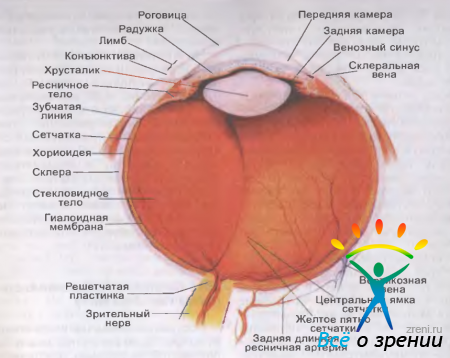
Small. 3.2. Budova full-time apple of the people.
In general, macroscopic eyes are seen, at first glance, we will forgive you: Two pockets (conjunctiva and one full apple) and three main shells (fibrous, sudinna, cyst), as well as the empty ones in the front and rear chambers (reserve with watery wolf), crustaceans. However, the histological structure of large fabrics is foldable.
The thinnest shells and optical centers of the eye are presented at the general distributions of the hand. The chapter is given to give the possibility of poaching the eye of the eye in general, the intelligence of the functional relationship between the parts of the eye and its appendages, the particularity of blood flow and energy, which will clarify the pathology of the changes in vision
Fibrous shell of the eye (tunica fibrosa bulbi)
Fibrous shell of the eye to be stored from rods and sclera, both in terms of anatomical structures and functional powers, one way of one is very different.
Rogivka (cornea) - the front opening is a part (~ 1/6) of the fibrous sheath. A simple transition to the sclera (limb) can be visually transparent and up to 1 mm wide. It’s clear that the little balls of the horns widen backward, trochi dal, below the front. Vidmіnnі strength of the horns: spherical (radius of curvature of the anterior surface ~ 7.7 mm, rear 6.8 mm), mirror-like blinding, blood-bearing vesicles are suppressed, there is a high tactile and painful, ale of low temperature, sensitive 43 diopters.
The horizontal diameter of the crotch in healthy newborns is 9.62 ± 0.1 mm, in older adults, the reach is 11 mm (the vertical diameter is smaller by ~ 1 mm). At the center of the won there is a subtle one, lower on the periphery. The whole indicator is also significant for the first time: for example, at 20-30 rockets the width of the flanges is 0.534 and 0.707 mm, and in 71-80 rockets - 0.518 and 0.618 mm.
When the walls are overwhelmed temperature of horn at limba the doorway is 35.4 ° С, and in the center - 35.1 ° С (at high critical temperatures ~ 30 ° С). In connection with cym in them, there is a possibility of growth of colored fungi with the development of specific keratitis.
Scho feel grabbing Then there are two paths: for the diffusion of the perilimbal vessel hedge, the anterior ciliary arteries, and osmosis with the front camera and the late birth.
Sclera - opacity of the part (5/6) of the outer (fibrous) shell of a full-time apple with a thickness of 0.3-1 mm. Vona is the most thin (0.3-0.5 mm) in the area of adequateness and at the point of exit from the optic nerve. Here, the inner balls of the scleri set up a gratuitous plate, the axons of the ganglion cells of the sieve pass through the yak, and they set up the disc and the stem part of the zorotic nerve.
The zones of stonewalling of the sclera are suitable for the infusion of an advanced internal vise (development of the staphyloma, the excavation of the disc of the healthy nerve) and factors, the first for all mechanical (subconjunctival growth in types of muscles, attachments Close to the stitching, the thickness of the scleri becomes 0.6 0.8 mm.
In the area of limba to see zlittya three absolutely different structures- horns, sclera and conjunctiva of an intramural apple. In addition, a zone is given as an important point for the development of polymorphic pathological processes - from igniting and allergic to swollen (papiloma, melanoma) and related to anomalies in development (dermoid). The limbal zone is richly vascularized behind the anterior vein arteries (throat arteries), which are 2-3 mm in the middle of it, but not only in the middle of the eyes, but at least in the middle of the arteries: Episcleritis and adjacent conjunctiva. According to the number of limba, the gossip is more dense and nerve-filled, and it is confirmed by short and short ciliary nerves. From a new entrance to the ticket, so to enter into the ticket.
Tissue sclera has few judgments, it may be more relieved of sensitive nerve endings and is subdued to the development of pathological processes characteristic of collagenosis.
To the surface of the scleri to hang 6 gummies... Besides, in niy є special channels (vipuskniki, emisari). Behind one of them, to the vertebral column, there are arteries and nerves, and after them, there is a venous storm of a pink caliber.
On the inner surface of the anterior edge of the sclera of sewing circular flute up to 0.75 mm wide. The posterior edge of the jogging knuckle protrudes in front of the spur, until the ciliary body is attached (the anterior knuckle attached to the sheath of the sheath). The anterior margin of the flute between the Descemet shell of the horn. The venous sinus of the sclera (Schlemm's canal) is located at the posterior edge at the bottom. The insha part of scleral death is occupied by a trabecular mesh (reticulum trabeculare).
Sudinna shell of the eye (tunica vasculosa bulbi)
Sudinna shell of an eye to be stored in three tightly knitted parts - a ridge, a wicker body and a choroid.
Raiduzhka (iris) - the front part of the sheath of the court, at the top of the two of the two, is not rosetted along the wall, but at the front, along the length of the surface, there is a disc shape with an opening (window) in the center.
On the edge of the zinitsa, a ring-like sphincter is formed, which is innervated by the pericuchial nerve. Radially, the dilator is innervated by a sympathetic nerve.
Ridge thickness 0.2-0.4 mm; there is especially a market in the root zone, that is, on the cordon with viykovy til. Itself here, with important contusions of the intramural apple, it can be її vidriv (iridodialys).
Viykovy (ciliar) tilo (corpus ciliare) - the middle part of the sheath of the court - to be located behind the iris, that is inaccessible to a bezposredny look. On the surface of the sclera, the ciliary body is projected at a 6-7 mm wide viglyad, which can be repaired at the scleral spur, i.e., at the appearance of 2 mm in the face. Macroscopically, two parts can be seen in a single circle - a plane (orbiculus ciliaris) 4 mm wide, between the toothed line (ora serrata) stencils, and ciliary (corona ciliaris) with a width of 2-3 mm, with 70-80 ciliaris ciliaris ). The dermal part of the ridge is approximately 0.8 mm in height and up to 2 mm wide and up to 2 mm wide.
The inner surface of the wicker body is tied with a crystal to the aid of the so-called vіykovy pasque(Zonula ciliaris), which can be folded without much thinner worm-like fibers (fibrae zonulares). Tsey girdle viconuє the role of the ring, to bring the kryshtalik. Win a ciliated cilia with a crystal in a single akomodatsіynі apparatus ochі.
Sudin on the edge to form for a rakhunok of two rear arteries (full arteries), which pass through the sclera at the posterior pole of the eye, and then go into the suprachoroidal space along the meridian 3 and 9 years; anastomosis is made of the reddened anterior and posterior short vein arteries. Sensitive innervation of a vyykovy body is the same as that of a ryadzhka, a ruchov (for the young ports of the amoodation) is a kind of okorukhovy and sympathetic nerves.
Chorioidea Above, the vermilion shell, whistling the entire posterior view of the sclera on the protrusion from the tooth line to the healthy nerve, pretends to be the posterior short vein arteries (6-12), which pass through the sclera at the posterior pole of the eye.
Horioidea can have a number of anatomical features:
- the sensitive nerve ends are relieved, so that the pathological processes do not occur in any pathological process;
- її sudinna hem does not anastomose with the anterior ciliary arteries, which, as a rule, when the anterior appearances are choroid, become intact;
- a great bed of judgment with a small number of double vessels (4 vortic veins) with a confidence in blood flow and a fall here in the early days;
- organically tied with a sieve, like when you become ill with a good idea, as a rule, it can also get involved in a pathological process;
- through the manifestation of the perіchorіoidal space, it is easy to see through the sclera. To settle down in a normal position in the main way to enter venous vessels, perforation in the area of adequateness. The role of stabilization is also played by the judges and nerves, which penetrate into the choroid from the wide open space.
The inner (sensitive) shell of the eye
The inner shell of the eye - сітківка (retina) - whistling in the middle of the entire surface of the vessel shell. In terms of structure, and hence, the function in them is two parts - optical (pars optica retinae) and vyykovy-iris (pars ciliaris et iridica retinae). Persha is a highly differentiated nerve tissue with photoreceptors, so it can take adequate light strips from up to 380 to 770 nm. The cya part of the shedding extends from the disk of the healthy nerve to the flat part of the vyykovy body, de end of the tooth line. Distant in the reduced to two epithelial spheres of the view, having lost the optical power, it curved the inner surface of the vyykovy til and the arch. Tovshchina of sits on small dilyankas: At the edge of the spinal nerve disc 0.4-0.5 mm, in the area of the foveolus with the gum line 0.07-0.08 mm, at the dentate line 0.14 mm. Before the piddle of the vertebral column, the sits are creeping in several zones: the bridle of the tooth line, near the disc of the healthy nerve and along the edge of the new beaches. On іnshіh dіlyankas іdnannya pukhka, so here it’s easy to see it from its own pіgmentnogo іnіtelіyu.
Mayzhe in every way optical part of the link store for 10 balls. Photoreceptors, turning up to pigment ingestion, are represented by cones (about 7 million) and sticks (100-120 million). The first ones are grouped in the central parts of the shell, others in the center of the day, and the maximum power is set at 10-13 ° East. Far to the periphery, the number of sticks gradually changes. The main elements of the sieve are located in the sturdy position of the vertically expanded support cells of Mueller and industrial fabrics. Stabilize the function of viscous and near-cord membranes of the sieve (membrana limitans interna et externa).
Anatomically, and during ophthalmoscopy in the city, two more important ones are clearly visible in the functional type of presentation - disk of the healthy nerve i Zhovta Plyama, The center of which is located at the 3.5 mm edge at the front edge of the disc. In the world of closeness to the jovti plexus of Budov, the suttas of the suttas change: a collection of spheres of nerve fibers, then - ganglionic cells, far - an internal plexiform ball, a ball of internal nuclei and pllexiform. Foveoli are represented only by a ball of flasks, which can be divided into separate buildings (the area of the central zone, which occupies the space of objects ~ 1.2 °).
parameters of photoreceptors
sticks: Dovzhina 0.06 mm, diameter 2 microns. The above-mentioned segments have a pigment - rhodopsin, which is a part of the spectrum of electromagnet light emission in the range of green spaces (maximum 510 nm).
cones: Dovzhina 0035 mm, diameter 6 microns. In three different types of cones ("chervonyh", "green" and "blue"), there is a healthy pigment with a variety of indicators of glinting light. In "red" flasks, vin (iodopsin) adsorbed spectral changes from the greens to ~ 565 nm, in "green" flasks - 500 nm, in "blue" - 450 nm.
Pigments of cones and sticks "woke up" in the membrane - discs of the first segments and integral white substances.
Sticks and cones Volodya bright light sensitive... The first function is to function when the mid-season is up to 1 cd * m-2 (low, cattle zir), others - up to 10 cd * m-2 (day, photographic zir). If the brightness rises between 1 and 10 cd * m-2, all photoreceptors function at the singing level (mesopic, mesopic zir).
Zorian nerve disc to be located in the nasal half of the sieve (at 4 mm from the rear pole of the eye). Because of the addition of photoreceptors, that in the field of vision is evident until the moment of the projection є the blind zone.
harvesting of sits WELCOME from two dzherel: a number of inner balls are recognized from the central artery of the sieve (inside), and the neuroepithelium - from the choriocapillary ball of the vascular sheath.
The veins of the central arteries and veins pass through the spheres of nerve fibers and partly in the spheres of ganglion cells. The stench is to make a sharuvata a capilary hedge, as there are no more in the foveoli with zhovtoy beaches (div. Fig. 3.10).
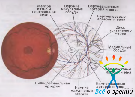
Small. 3.10. Topography of the central arteries of the central arteries and the veins of the eyes of the left eye on the diagrams and photographs of the face-to-face bottom.
The important anatomical peculiarity of the cells is those that are the axons of the ganglion cells on the whole added metal cover(One of the factors, which is the beginning of the tissue penetration). In addition, there, like a sheath of the ship, is relieved of sensitive nerve endings.
Internal core (empty) och
Porozhnin was able to take revenge on the light-conducting and light-refracting centers: the watery vologue, who remembers the front and back chambers, the crys- talik and the tough ones.
Anterior camera ochi (camera anterior bulbi) is a space, enclosed by the rear surface of the horn, the front surface of the ridge and the central part of the front capsule of the crystal. Misce, the derogivka go into the sclera, and the ridge - into the vіykovy tilo, called the cut of the anterior chamber (angulus iridocornealis). In the first place, there is a drainage (for a childlike vology) system of eyes, which is stored from the trabecular mesh, the scleral venous sinus (Schlemm's canal) and collector canals (vipusnikiv). Through the window, the front camera can be viewed from the rear. In the whole mission, there is a lot of glibin (2.75-3.5 mm), which step by step Change from right to the periphery (div. Fig. 3.2).
Rear camera of the eye (camera posterior bulbi) to be located behind the iris, like the front wall, and surrounded by a warm tone, behind a spiteful tone. The internal wall is set up by the kryshtalik's eqvator. The entire space of the rear chamber is permeated with the clinks of a vyykovy pasque.
At the norm of offended cells, the eyes are filled with a watery vologue, as behind their warehouse there is a dializate of blood plasma. The watery vologue should take revenge on lively speech, winter glucose, ascorbic acid and acid, which can be consumed by crystal and horny, and taking away from the moment the products are exchanged - lactic acid, carbonic acid
The offense cameras of the eye accommodate 1.23-1.32 cm3 of a line, which becomes 4% of all eyes. Chilinny obsyag chamber vologue in the middle 2 mm 3, dobovy - 2.9 cm3. In other words, a new exchange of chamber vologists will take 10 years.
Between flooding and current internal flow equally important balance... There are some reasons for failing, and it is necessary to create an internal vice to the point of changing the level, the upper boundary in the norm does not change 27 mm Hg. (When vimіryuvannі with a Maklakov tonometer weighing 10 g).
With the main ruinous force, I will get an uninterrupted strum of the line from the rear chamber to the front, and then through the kut of the front chamber between the eyes, є gap in empty eyes and venous sinus sclera(Close to 10 mm Hg), as well as in the designated sinus and anterior veins.
Krishtalik (lens) a semi-solid no-court body in the form of a double-convex linseed, laid in a capsule, with a diameter of 9-10 mm and a thickness (in the form of storage) 3.6-5 mm. The radius of curvature of the front surface in the quiet adaptation of the road is 10 mm, the rear - 6 mm (at the maximum load of the adaptation of 5.33 and 5.33 mm, it seems), that in the first fall, the breaking force of the middle 19 is to become in another 33.06 diopters. In new-born women, Kryshtalik may have a lot of kulastiy, and have a soft consistency and breaking strength up to 35.0 diopters.
In the otsi kryshtalik is located immediately behind the ridge in nasty yamts(Fossa hyaloidea). In most cases, wines are reduced by numerical gum-like fibers, which in the sum is set up with a girdle (vyykovy girdle).
The back surface of the kryshtalik, so the front surface itself, is washed with a watery vologue, splinters on the whole face are lifted from the obscene body by a vuzkoy schilina (retroletal space - spaiium retrolentalc). However, along the last edge of the insect-like fossa, there is a space between the lower ring-like ring of the Viger, the rosette between the crystal and the middle-class body. Kharchuvannya krishtalik go along the path of exchange processes from the chamber vologo.
Camera vitrea bulbi the rear view of the empty space is filled with the closest type (corpus vitreum), like the front of the bed up to the crystal, which is set in a small space (fossa hyaloidea), and on the sieve It is a hole in a dralist mass (like a gel) with a volume of 3.5-4 ml and a weight of approximately 4 m. However, only 10% of water is tied with the components of a sparing body, so the exchange of a line in a new one should be reached actively and at a distance, for deyakim data, 250 ml for extra.
Macroscopically, they see the vascular stroma (stroma vitreum), which pierces the occlusive (Cloquet) canal, and the navkolishny call of the haloid membrane (Fig. 3.3).
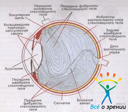
Small. 3.3. Inexperienced people (sagital sphiz) [according to N. S. Jaffe, 1969].
sclopodibna stroma store up to fill up the fluffy central speech, in which є optically empty zones, filled with ridinoy (humor vitreus), and collagen fibrils. Remain, narrowly, set up a number of real paths and a large cortical ball.
hyaloid membrane to be stored in two parts - front and rear. The cordon between them to pass along the tooth-like line of the sieve. At its side, the anterior near-cord membrane has two anatomically mounted parts - crystalline and zonular. The cordon between them is Viger's circular haloidocapsular connection. mіtsna tilki in a childish vіtsі.
In the area of the so-called front and rear seats, they are not tied to the side. After the first size of the area, it’s not easy to get it immediately until the end of the day is 1-2 mm in advance. scalloped edge(Ora serrata) stitches that are 2-3 mm behind. The back of the mucous membrane is the zone of fixation of the thymus near the spinal nerve disc. Vvazayut, sclopodіbne tіlo maє links with sіtkіvkoyu also in the area of macula.
Sklopodibny (Cloquet) canal (canalis hyaloideus) funnel-like enlargements from the edges of the healthy nerve disc and pass through the stroma right up to the posterior capsule of the crys- talik. The maximum width ka-n & pa is 1-2 mm. In the embryonic period, in the new, the artery of the obscene til passes through, until the moment of the nation's life the child is deserted.
The yak has already begun, in the wicked tili іsnu persistent strum of rіdini. From the rear chamber of the eye of the bed, which is produced by the wicker body, through the zonular slot, it is consumed in the anterior wedge of the bedrock body. Dalіdina, scho poured in the bed, collapses to the squeeze and prepapillary opening in the hyaloid membrane and emerges from the eye through the structures of the healthy nerve, so along the perivascular spaces of the retinal vessels.
Zoroviy path and path of the pupillary reflex
The anatomical structure of the zorovaya pathway is foldable and includes a number of neuronal lanes. Between the eyes of the skin eye - tse
- ball stick and cone (photoreceptor - I neuron),
- small ball of bipolar (II neuron)
- and ganglionic cells with other axons (III neuron).
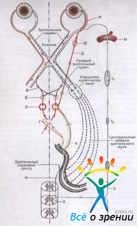
Small. 3.4. Zorovi and pupils of the way (diagram) [after S. Behr, 1931, in zmіnami]. Explained in the text.
Healthy nerve (n.opticus) statements by axons of ganglionic cells of the sith and end in chiasm. In older people, the size of the gale is varied from 35 to 55 mm. I mean a part of the nerve to become an intra-hollow vidrizok (25-30 mm), which is an S-padded vigin in a horizontal area, which does not see tensions when an intramural apple collapses.
On a significant projection (from the entrance from the intramural apple to the entrance to the healthy canal - canalis opticus), a nerve, similar to the brain, maє three shells: Solid, pavutinnu and myaku (div. Fig. 3.9).
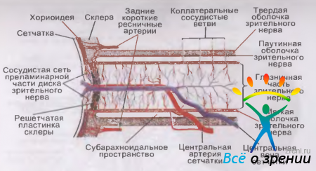
Small. 3.9. Blood posture of a zorovy seal and a sieve (scheme) [according to N. Remky, 1975].
At the same time, the thickness should be 4-4.5 mm, without them - 3-3.5 mm. In an intramural apple, a hard cerebral sheath grows in the sclera and tenon capsule, and in the canal in the canal - in the oyster. The intracranial nerve and chiasma excrescences, which are located in the subarachnoid chiasmatic cistern, are drawn into the pulp sheath.
Intracheal space of the intracollective part of the nerve(Subdural and subarachnoid) to be confined to the analogous expanses of the brain, albeit isolated one from one. The stench is stored in a folding fold (intraocular, tissue, cerebrospinal). Oskilki internal vise in the norm is 2 times as in the internal cranial (10-12 mm Hg), straight into the struma to form a vise with a gradient. The winch becomes vypadki, if the internal cranial grip is frequently moving (for example, during the development of puffiness to the brain, bleeding into the empty skull);
All nerve fibers that enter the fold of the healthy nerve, group in three main beams... Axons of ganglionic cells, which enter the central (macular) region of the sieve, fold the papillomacular bundle, which enters the skrone half of the zorotic nerve disc. Fibers from ganglionic cells of the nasal half of the sieve travel along radial lines into the nasal half of the disk. Analogous fibers, ale from the rim half of the sieve, on the way up to the disk of the healthy nerve from above and below “wrap around” the papillomacular bundle.
In vіdrіzku The healthy nerve, close to the intramural apple, will be filled with the same nerve fibers as in the disk. Distant papillomacular bundle moves in the axis of position, and the fibers from the fringe quadrants of the suture - to the entire half of the green nerve. In such a rank, the healthy nerve is clearly divided on the right and left half. The mensch is twisted in the upper and lower halves. Important in the classroom sensation is special є those who are the nerve of the release of sensitive nerve ends.
At the empty skull Good nerves to settle over the area of the Turkish saddle, fixing chiasma (chiasma opticum), the yak is covered with a soft cerebral sheath, and the size of the size is 4-10 mm, width is 9-11 mm, thickness is 5 mm. Chiasma from the bottom between the diaphragm of the Turkish seat (the duralum of the dura mater is taken up), from the top (in the back side) - from the bottom of the third slit to the brain, from the sides - from the inner sleepy arteries, from the back.
In the area of hiasma the fibers of the healthy nerves are often crossed over the rakhunok of the ports, tied with the nasal halves of the retinas. Go over to the opposite side, smell the fibers, go out of the skinny halves of the retinas of your eyes, and make the paths look good. There and then partly overlapping і papillomacular bundles.
Zorovi tracts (tractus opticus) to repair at the back surface of the chiasm and, bending around the lower side of the brain in front of the brain, to end in the newer side (corpus geniculatum laterale), the back part of the corpus of the green hillock (thalamus side of the opticus) However, it is only a zvnіshnі kolіnchati tіla є a crazy young green center. Two reshta to highlight the functions.
In healthy tracts, more than 30-40 mm, the papillomacular bundle also occupies a central position, and crossed and uncrossed fibers and earlier go in close bundles. At the same time, they roztasvani ventromedial, and others - dorsolateral.
good radiance(Fibers of the central neuron) to repair from ganglion cells of the fifth and first balls of the most frequent body. A collection of axon cych cells is set up in the so-called Vernike field, and then, passing through the posteriorly inner capsule, it vially likewise diverges into the bile speech of the cerebral part of the brain. The central neuron ends in a bird spur (sulcus calcarinus). Qia region and a sensory healthy center - 17th cortical field according to Brodman.
Shlyah pupillary reflex- svitlovogo і for setting the eyes on a close view - to finish folding (div. Fig. 3.4). The afferent part of the reflex arc (a) of the first of them is repaired from the flasks and sticks of the sieve at the view of autonomous fibers, to go to the warehouse of the healthy nerve. At the chiasm, the stinks are crossed in the same way, like the healthy fibers, and go to the healthy paths. In front of the new collocated bodies of the pupillomotor fiber, there is a drop in the brachium quadrigeminum, or the so-called area pretectalis. Distant new, industrial neurons for partial crossing are directed to the external nuclei (Yakubovich - Edinger - Westphal) of the okoruchial nerve (c). Afferent fibers from the same membranes of the cutaneous eye stitch are presented in both okorukhovy nuclei (d).
Efficient way of innervating the sphinkter of the Rayduzhka to repair from the already missed nuclei and from the emerging beam in the warehouses of the okoruhny nerve (n. oculomotorius) (e). In the inner pit, the sphincter fibers enter into the lower head, and then through the cortex (radix oculomotoria) - into the ciliary vuzole (e). Here the first neuron of the outgoing path will end and the other will be repaired. When the fibers of the sphincter are sent to the warehouse of the short chain nerves (nn. Ciliares breves), passing through the sclera, they enter the perichoroidal space, making the nerve gossip (f). Yogo kіntsevі rozgaluzhennya penetrate into the іnuzhka and enter into the muzzle with only radial beams, that is. In total, there are 70-80 such segments in the business manager.
Dilatator pupillae , I will take off my sympathetic energy, I will try to go to the central center of Budge. The rest is located in the anterior horns of the spinal cord (s) between the SCC and ThM. Svidtsi enter from the center of the sympathetic nerve, like through the near-cordonny stub of the sympathetic nerve (l), and then from the lower and middle sympathetic ganglia (t, and t2) reach the upper ganglion (t3) (CIV). Here the first neuron of the path ends and the other is repaired, to enter the storehouse of gossip of the internal sleepy artery (m). At the empty skull, fibers, which innervate the dilator of the brain, go through the guessed gossip, enter the three-part (Gasser) vuzol (gangl. Trigeminale), and then fill it in the warehouse of the intravenous nerve (n. Ophthalmicus). Already at the apex, the ochitic stench passes into the nasociliaris nerve (n. Nasociliaris) and at once through the ciliary nerves (nn. Ciliares longi) penetrate into the ovary apple.
The regulation of the function of the dilator of the brain is directed to the auxiliary supranuclear hypothalamic center, which is located at the bottom of the third brain in front of the hypophysis. For an additional form of dressing wines from the Budge center.
The reaction of the brain to the convergence and the adaptation of one's own particularity, and reflex arcs in the whole type of vidrіznyayayutsya in the description of the food.
With the convergence stimulus to the sound of the zinitsa, to serve as proprioceptive impulses, so that the inner straight mouths of the eye go to speed. Accommodation and incentives to dispense (focus) The eferent part of the arch of the pupillary reflex is the same in both cases.
The center of setting eyes on the close is located, yak vvazhayut, in the 18th cortical field along Brodman.
Ochnitsa (orbita) і її vmist
ochnitsaє kistkovym vstylische for a full-time apple. Through the empty, back (retrobulbarny) formations of fatty body (corpus adiposum orbitae), pass the healthy nerve, ruffs and sensitive nerves, okruchs, muses, fascial vernaculars, Skin ochnitsa has a form of truncated chotirigranous pyramid, with a brutal top in the side of the skull at 45 ° to the sagittal area. In the growth of a human, the height is 4-5 cm, the horizontal diameter of the entrance (aditus orbitae) is close to 4 cm, the vertical diameter is 3.5 cm (Fig. 3.5).
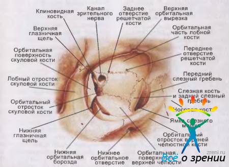
Small. 3.5. Ochnitsa (right).
Three of the chotiroh stinok ochnitsi (krym zovnishnyoi) mezhuyut from the nasal sinuses. It is not easy to serve as a sinister reason for the development in these quiet pathological processes, a bowl of fiery nature. Possibly and sprouting chubby, how to go out from the thick, frontal and upper slit sinuses.
call, Nybіlsh mіtsna і naymensh is inflamed in case of illnesses and injuries, a small part was approved by a wedge, partly by a lobe and a great krill of a wedge-like wedge. Tsya stinka vidokremlyu instead of ochnichi from the early fossa.
Upper glass ochnytsi molded in the main frontal wedge, in comrade, as a rule, є sinus (sinus frontalis), and partly (in the rear end) - malim krill wedge-like wedge; Between the anterior cranial fossa, and the tense surroundings, the severity of young people begins to accelerate in times of difficulty. On the inner surface of the intra-well part of the frontal knuckle, at the lower edge, there is a small knuckle knuckle (spina trochlearis), up to which a tendon loop ripples. The tendon of the upper oblique muzzle passes through it, as it travels straight ahead. At the upper-outer part of the frontal cyst, there is a fossa of a lacrimal zoloz (fossa glandulae lacrimalis).
Internal wall ochnitsi in the great country it was approved by the even thinnest cyst payment - lam. orbital is (raruracea) gratuitous kistki. From the front to it, there is a fold with the rear lacrimal comb and the frontal extension of the upper gap with the front lacrimal comb, at the back - just a wedge-like fold, at the top - a part of the frontal fold - a part of the frontal fold, Between the combs of the lacrimal brush and the frontal protrusion of the upper slit sl_zniy m_shok(Saccus lacrimalis). At the bottom, the fossa passes into the nasolacrimal canal (canalis nasolacrimalis), which is located in the upper slit cyst. Win revenge nasolacrimal duct(Ductus nasolacrimalis), which ends at a distance of 1.5-2 cm behind the anterior edge of the inferior nasal concha. After all, it’s easy to navigate with blunt injuries with the development of emphysema capital (partly) and self-centered (older). In addition, the pathological processes that occur in the cracked grooves, end up widening towards the ochin, as a result of which the ignition block of my tissue (cellulitis), phlegmon or neuritis of the green nerve develops.
Bottom wall ochnytsiє one hour and the upper wall of the upper slit sinus. The cya stinka was approved as the head rank of the vernacular surface of the upper slit, sometimes also the vilified cyst and the orbital with the small pedunculate cyst. In the case of a dangerous person, there may be fractures of the lower wall, which are sometimes superimposed on the lowering of the intramural apple and the interfering of its collapse, the name is when the lower oblique meat is interconnected. To fix the lower part of the ossicle from the cyst, the trochus lateral to the entrance to the nasolacrimal canal. Firing and plump processes, which develop in the upper slit grooves, can easily expand towards the ochnitsa.
At the summit, in the walls of ochnitsa, a number of openings and cracks, a number of great nerves and blood-bearing vessels pass through the empty slopes.
- Cystkovy canal of the zorian nerve(Canalis opticus) up to 5-6 mm. To repair in an ochnitsa round opening (foramen opticum) with a diameter of close to 4 mm, from one empty to the middle cranial fossa. The zorovy nerve (n. Opticus) and the intra artery (a. Ophthalmica) enter through the canal into the vascular fossa.
- Upper orbital cleft(Fissura orbitalis superior). Established by a wedge-like cyst and krils, I will close the fossa with the middle cranial fossa. Tightened by a swampy, semi-tissue rye, through the yak into the intramural fossa, three main glands of the intramural nerve (n. Trochlearis, abducens and oculomotorius). Through qiu zh її її leaves the upper internal Vienna (v. Ophthalmica superior). With the development of a characteristic symptom complex: ophthalmoplegia povna, i.e. immobility of the intramural apple, drooping (ptosis) of the upper line, mediasis, decreased vision of dyspepsia and malformations However, the "syndrome of the upper orbital fissure" may not be rotated, if not all of them, but if not all of the nerves start to pass through the fissure.
- Lower orbital cleft(Fissura orbitalis inferior). The bottom edge of the great krill of the wedge-like wedge and the trough of the upper slit is fixed, which will prevent the formation of eyebrows from the wedge-like (in the back half) and the lower fossa. The tsia shchilina is also covered with a semi-tissue cross-flow, the fibers of the orbital mucosa (m. Orbitalis), innervated by a sympathetic nerve, are embedded in the yak. Through it, the fossa is filled with one of the two holes of the lower intravenous vein (which falls into the upper intravenous vein), which anastomoses with the cryopodic venous plexus (et plexus venosus pterygoideus), and enter the lower nerve arterial venous Zygomaticus) and intramural cells of the cryoplastic university (ganglion pterygopalatinum).
- round hole(Foramen rotundum) is located near the great wedge-like krill. I will tie the middle cranial fossa from the crylopidnebyno. Through the end of the opening pass another tricycle nerve (n. Maxillaris), from which the infraorbital nerve (n. Infraorbitalis) enters the crylopidnebine fossa, and the ciliated nerve (n. Zygomaticus) into the inferior temporal. A nerve injury then penetrates into the empty ochnitsa (subperiosteal perspiration) through the lower ochnoyamkovu crack.
- Open a part at a medical school(Foramen ethmoidale anterius et posterius), through which one nerve passes (nasal nerve head), arteries and veins.
In addition, in the great krill wedge-like cyst, there is one more opening - the oval (foramen ovale), where the middle cranial fossa is located from the middle of the cranial fossa. The third branch of the tricyclic nerve (n. Mandibularis) passes through it, but you don't take part in the innervation of the organ.
For a full-time apple on the side of 18-20 mm from the rear pole there is eyelash vuzol(Ganglion ciliare) with a size of 2 x 1 mm. The wrinkles are folded down to the surface of the healthy nerve. Vykovy vuzol - peripheral nerve ganglion, cells of which behind the other three roots (radix nasociliaris, oculomotoria et sympathicus) are tied with fibers of the external nerves.
Quite a few pictures of fine, ale motsnoy around(Periorbita), which is naturally grown up with them in the area of the cyst sutures and the green canal. The openings of the rest of the tendon ring (annulus tendineus communis Zinni), from which all okorukhs are repaired, behind a vignette of the lower scythe. Vona grabbed a cob from the lower cistern of the ochnitsa, near the entrance to the opening of the nasolacrimal canal.
Outside, up to the fascias, according to the International Anatomical Nomenclature, one can see the apple, the fascia, the orbital septum and the fatty of the intracellular fossa (corbitus adiposum).
Pichva full-time apple (vagina bulbi, colishnya name - fascia bulbi s. Tenoni) pokrivaє mayzhe everything is very yabluko, behind a vignette of a horn and a misce to the entrance of the zorovy nerve. The greatest degree of competence and development of the fascia is in the area of adequate eyes, where the tendons of the mucous membranes pass through it on the way to the point of attachment to the surface of the sclera. For a measure of closeness to limbu fabrics, they groan and at the end of the day, step by step, get involved in the subconditions of fabrics. In people with extraocular muses, you can reach a thin connective tissue coating. From the center of the zone to enter the thick (fasciae musculares), so that the eyes ring from the sides of the glasses and the edges of the eyes. As a whole, they form an annular membrane, which is parallel to that of the eyes and fixes it in the internal pit in a stable position.
Subvaginal space of the eyes(The colishnya is called spaiium Tenoni) is a system of clefts in the fluffy episcleral tissue. Vono will get a wild apple in the singing society. A lot of space is not easy to pick up with surgical and therapeutic methods (sclero-strengthening surgery of the plantation type, introduced by lykarsk zasobіv by way of іn'єktsіy).
orbital septum(Septum orbitale) - the structure of the fascial type is kindly rotated, it is roasted at the frontal area. Along the side of the cartilage of the capital, with the cystic edges of the bone. At once, the stench will make you feel like a bite, a crumbling, a stink, like an empty ochnitsa when the world is darkened. It is important to pay attention to the fact that in the area of the medial stage there is a good partition, I also call it Tarzi-orbital fascial, creep up to the back lacrimal crest of the lacrimal fold, in case of someone else, it’s easy to know what is next to it. . the pose of an empty ochnitsi.
Empty blanks are stored fat body(Corpus adiposum orbitae), which is placed in a thin aponeurosis and penetrated with semi-tissue loops, which can be found in other segments. The rules of plasticity of adipose tissue do not overtake the ability to pass through its okorukhovym (in case of fast) and healthy nerve (in case of collapse of an intramural apple). From the fatty tissue it is adorned with a wedge-shaped space.
Through a vomiting hole, straight from the top to the entrance, pass through the blood-bearing judges, rukhovs, sensitive and sympathetic nerves, as well as a little bit of viscera, and a report at the head's head. The same goes to the healthy nerve.
Continued on the offensive stat: Is the anatomy normal to the organ of a human being? Part 2