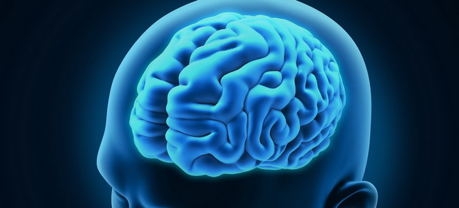
Sagittal development of the brain
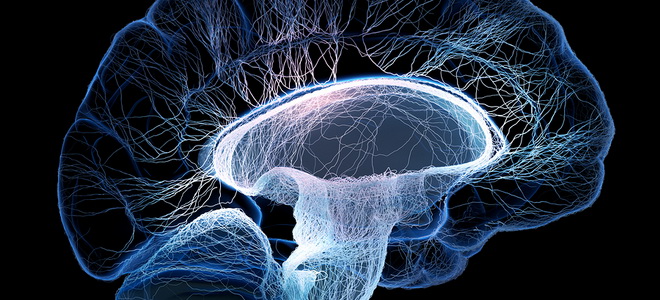
Understanding about the sagittal development of the brain has a key role in the scientific growth and function of the organ, diagnostics of the development of diseases and the predictable stems of the disease. Sagittal growth - a chain of strong lines, yak, passing through the empty brain, to extend it to the right and left of the part. This sagittal line makes it possible to integrate the vision from us with storage components that are accessible to the eye.
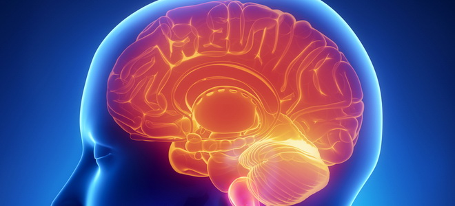
The brain in the form of a post is composed of some of the main components:
- Kintseviy or the great brain is the most important part of the organ, the yaka hangs over the first dilyankas
- The industrial brain - practical ghosting of the eyes in the great comrade
- Moss stovbur with a dovgastim brain, a bridge and a middle brain, which extends from the bottom to the top of the dorsal cord at the view of the cylindrical cord. Є with a good lanka between the head and the back
- The lobe is a small tree-like light, it is clearly rooted in the back of the brain
The most significant part of the nervous system is the endometrium, the function of which is responsible for the coordination of the robotic arm and the spinal cord. We look for a regulator of nervous activity є the cortex of the brain, like a cloak covering the surface of the great brain.
On the growth of the cortex, there is a yak ball of dark fabric, from 1.5 to 4.5 mm in size, clinging to the brain. There are a lot of nerve cells, which are close to 12 - 17 billion, skin from several thousand to tens of thousands of contacts with small cells. Tsі klіtini roztashovanі pozharovo in 6 wіddіlіv and get in touch with the foreign minds of cytoarchitectonics:
- Molecular ball with a small number of nerve cells and numerical tangles of nerve fibers
- Significant granular ball of neurons of small sizes in the form of granules
- The newest ball from the middle and the middle for the size of the nerve cells in the form of pyramids
- Internal granular ball, which is close behind Budova to another ball
- Internal pyramid ball with giant neurons behind the size of the pyramid
- Multifaceted ball, which can be stored from nerve cells in the form of spindle and tricycle
The cytoarchitectonics of Bula was described by the great Russian scholar - I.P. Pavlovim. On the other hand, the six-ball system of visualization plays an important role in the conducted nerve impulses and responses. If you fix it with a part, the pulse will end without the middle, moreover, the skin will feel like it will show its cytoarchitectonic field.
In addition to the ordered structural ball-like shaping of nerve cells, these growths can also have a consistent pattern, explained to the understanding of myeloarchitectonics.
Nerve fibers, spreading six balls, go horizontally, intertwining with vertical bundles of nerve spikes. I will arrange such a type of nerve nobles, allowing the robot to coordinate the brain like in the middle of measles, so for the help of the root of the ring behind the boundaries.
A great brain, which is built up from two pivots, separated by a late furrow, on the basis of a post at one viglyad - either the left or the right. Pivot to be stored from the decal, to merge one into one surface - the upper lateral, which is borrowed from the upper and the most common, to the bottom of the brain, medial, like to be cultivated in the late late pіvkulі, and th lay down to them the bottom of the brain іddіli.
I will use a spike between myself and a spike - calluses with a little bit of calluses, as half of it is consumed in the eyes.
Skin infusion of the brain may have a folded ridge, for example, the surface of the grooves and zivin were streaked on the surface, which can be attributed to:
- First - the most glib, early and permanently present in the brain
- Secondary - this is also often the case, however, they are less significant than the first
- Tretinni - non-persistent and minlivi
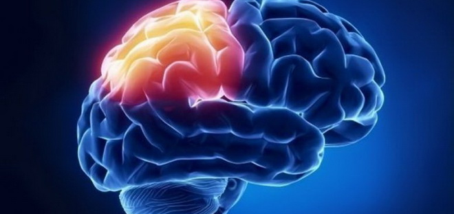
Behind the help of the primary grooves, the drink lasts for 5 parts - lobova, tim'yanu, skronev, potilichna and insular:
- Lobova share, scho zalyagaє in front of pivkul brain. Tsia dilyanka is promising for the message of comprehension of rukhiv, for the function of moving and misrepresentation, as well as for the formulation of non-standard forms of behavior
- This part of the house grows to be located at the upper bicnaya part of the drink and the projection center of sensitive views on human organism. Vaughn the projectє tactile vision, feeling the vagi і double-minded sensitivity, seeing the volume of rukhiv and myazovo-sloping pose
- Skrone part of the loan the lower lateral view of the brain and organizing savor, rumors and scent messages, as well as taking care of the sound of moving, mental performance and memory
- The strong part is localized in the rear part of the brewery and it is practically not seen as a temporary and early part, cordon between the zones of rubbing. It is important for the brain to keep track of key information as to how healthy organs are.
- Ostrіvkova part, very close to one of the grooves in the brain, busy with the analysis of savory views
Stovburovo іddіl to the brain
Stodburr brain includes a small brain - light in the form of a truncated cone is close to 25 mm in length, as it stretches directly out of the spinal cord from the bottom and there are little furrows with it, and its upper part is dragged by the main part.
It seems to be seen from the bottom and the top cordon. The lower I bet of the spinal nerves on the rivna of the great opening in the country. The upper and lower edge of the bridge.
The front surface should be avenged on the pair of children - pirudin and nerve fibers are tied clearly with them, in front of which the structure of the oval shape - the olive to the brain - is formed.
The statements made between the piracy and the olive canal serve as a motto for the origin of the two cranial nerves - pid'yazic nerve. Behind the Olivia lie nine, ten and eleventh pairs - the lazyopharyngeal, bloody and pre-cranial cranial nerves.
On the rise, it can be seen that the brain becomes gray and white. Microscopic dosage is given in respect of the housekeeping, so that the sera speech is stored okremi, in shape and size of a group of nerve cells - the nucleus of a dense brain.
The posterior viddil includes the nuclei of the twelve, eleventh, tenth, ninth and eighth pairs of cranial nerves - pidyazic, dodatkovy, bloody, lingopharyngeal and frontal-ultkovy.
In addition to the nuclei of the cranial nerves, to the fold of the large brain, enter the switching nuclei - a massive group of cells of the growth form. Nybilshi - olivia kernels - light in the form of eelpine up to 10 mm, which grow in the olivine brain and form the industrial center of the rivnova. In addition to the core, which is responsible for the formation of a high and partial tactile sensitivity, as well as painful and temperature perception.
The core of the nuclei, in the central part of the syllable of speech in the deep brain, is covered by the name of the reticular formation, as it is stored in the form of mutually reinforced cells of nerve cells.
The whole component plays an important role in the control of the small parts of the central nervous system, it accelerates the spinal cord reflexively, as well as takes care of the fate of the measles tonus of the great diseases. In a wide speech, nerves and fibrous bundles are presented, so that they pass through the brain through the structures.
The upper dilenka of the large brain passes into the varolyus mist - a structure, which is carried to the stovbur and roztashovanu in front of the middle brain. Dilanka of the bridge, the beasts backward, fixing the brainwash - a quarter-hole to the brain, or a diamond-shaped pit.
A tripartite nerve passes through the side of the bichesky surface of the bridge. Mіst at once with a dovgastim brain and cerebellum set up a kut, which I will call the cerebellopontine. Tsei kut є mіscem passing through the cranial nerves.
The brain is allowed to see the front and back of the bridge in the bridge, which I call the basilar part and the cover of the bridge. Rozdіlyaє їkh cordon to serve as a trapezoidal tilo. Yak і dovgastiy mozok, the place to be built up from the bіloї і syroї speeches.
The step behind the bridge is seen in the middle of the brain - the middle brain, which moves in front of the brain. Head storage parts of the middle brain є nіzhki і dax. Two great rollers-like illumination, which are stored from the later expanded nerve fibers, make up the brain.
Roztashovani tsi strukturi gostrym kutom and mute the middle hole, from closing it with a thin plate, as we call it a rhyme with speech. It is tied with a tse z tim, so the whole poevrhnost is tackled with openings to enter and go out of blood-bearing vessels.
The back surface of the middle brain is whistling, yak is inserted into the brain with great infusions. The cone-like zalosa dax grows to the upper and lower part, represented in the viglyadi humps.
From them, reach out to illuminate the eyes of the handles, so that the middle brain is drawn from the middle. Internal in the middle of the brain є a small channel width, which borrows up to 2 cm in a total - water supply to the brain. Its role is to establish the ringing of the third and fourth licks between oneself.
In the middle brain, the anterior and posterior surfaces are. Behind the rosette, wear the name pokrishki, store with a complex of white and syroi speech, and in advance - present, store with white speech. Between the tsimi parts, the chorna substance is deposited from the chorny pigment melanin.
Tsya dilyanka to the brain is coordinated to the robot of myazovaya tone, and also to follow the folding rukhovny acts, such as yak zhuvannya and kovtannya. The middle brain is indicative of reflexive rumors and healthy acts, which are based on ruffian, emotional and defensive reflexes on light and sound imitations.
Intermediate brain and cerebellum
Back from the cerebral stub, the brain grows, but it does not change 150 m. Yak and great brews, divided by schilines and bearded into pieces, leaves and pieces, and on the surface.
With a gray speech in a corn, I pretend to be a surface of the trisharov's bark, which I used to dry in my nads. Three balls, which whistle the bark, are represented by light ring molecular, middle pear-shaped and dark inner granular.
In the anatomical plan, the stovbur is replaced by the midbrain. Phylogenetically, it is important to breed for older and younger children. Up to the number of young people who are familiar with the understandings - the thalamic brain, carry the thalamus, metathalamus and epithalamus, and before the elderly the hypothalamus.
The head thalamic illumination is the thalamus, in the viglyad of the paired great egg-like speech it grows in the brain. The yogo back edge of the pleasures is called a pillow. The offense of the thalamus is from 'dnanі zroshchennyam, as I can call it interthalamic. The growth of the brain shows that the thalamus is one of the most hard-pressed purchases of syroi speech.
Thalamus is an important center for processing sensitive visions. Until new signals come, impulses from the receptors of the shkiri, the vestibular apparatus of the internal vuh of the people, the musculoskeletal organs.
Information from healthy and auditory analyzers is transmitted to the metathalamus. The whole trail of sensitive visions, which passes through the thalamus, straight into the bark of the great pimples - the food center of the processing and analysis of information.
The thalamic nuclei are straightened to the stem of the optimal quick and short-hour reaction for the specific situation, which is a winicle. Hypothalamus, disassembled immediately before the thalamus, can be stored in more than 30 pairs of nuclei in its warehouse, which can be stored in three groups - anterior, middle and posterior. The hypothalamic nuclei establish great neuronal connections with the other structures of the brain - the thalamus, the lambic system, the hypophysis and the reticular formation.
Lingers to the brain
The brain has є empty, filled with cerebrospinal fluid. When empty, they sound like shlunochkiv. In total, there are 4 little ones - bichni - right and left, as they show their skinny and third to fourth.
Bichnі shlunochki prikhovanі pіvkuli kіntsevoy marz і may be the central vіddіl with three horns - forehead, tilted і skroneviy, skin from which can be directly seen, as seen in parts. Bichnі shlunochki behind an additional opening, z'єednіy with the third shlunochku.
The third slumber has three walls - the anterior from the end plate and the anterior adhesions, the posterior from the tip of the pineal body and the upper, from the callus to the top, and the nemov dakh curled the slumber from the top. In the coverage of the fourth slumber, the role of the dovgastia cerebral, mest and cerebral plays a role.
The whole creeper is stored from the bottom, from the bottom, from the bottom, from the bottom. At the bottom, I see a diamond-like fossa, lined with lobes. The upper part of the fossa comes from the aqueduct of the middle brain, and the lower part from the central canal of the spinal cord.
The dax of the fourth ditch was approved by the upper cerebral vitril from the plate of the white speech in the form of a tricot, the speech of the corn and the lower cerebral vitril from the embryonic surplus of the rear part of the primary microchur to the brain. Behind an additional row of empty openings, the lid is tied to the subarachnoid space of the brain.
Sagittal development of the brain gives an indication of this budget in the picture, allowing to assess the structure of the brain, without breaking the integrity of the internal warehouse. So, as the offense to the brain appears to be practically identical to the mirror, the sagittal development of any of them is given a sketching information about the structure of the brain.
In addition to scientific goals, tied to the implantation of anatomical features of the organ, the development of a clear understanding of the vast interposition of structural components in the brain.
The price is less important in the neurosurgeon's robots, who is guilty of volodia in general information about the exact storage and localization of the brain. Determining the development of the brain of a patient who has been rejected by the method of diagnostic methods for the type of MRI, which is based on the ballistic results before the diagnosis, with normal sagittal development of the brain, it is possible to draw an analogy and strikingly.