Wonderful for the "Head brain" in the dictionaries:
Brain
Brain (Encephalon)(Fig. 258) roztashuutsya in the empty cerebral skull. The middle wall of the brain of a grown-up human becomes approximately 1350. The wine has an ovoid shape through the protruding frontal and longitudinal poles.
On the last lump of the upper lateral surface of the brain (Facies superolateralis cerebri) to grow the number and size of the furrow (Sulci cerebri)(Fig. 258). Above, do not go into them, there is a pavutin of the sheath of the brain. From the poles of the pole to pass the transverse slit of the great brain, before which is the bed of the brain, which is the most important center for the coordination of the arms. For the middle line of the brain, pass the later day (Fissura longitudinalis cerebri), І і і і іve pіvkuli (Hemispherium cerebri dextrum et sinistrum)... Lower surface (Fasies inferior cerebri) characterized by a folding top.
At the empty skull, the spinal cord is trivialized with a gas-stimulus, which can be used to take revenge on the vertebral cord and dichal centri. The growth and growth of the lower part of the brain and the brain is connected one by one behind the auxiliary bridge, which is removed from the large brain. The lobe grows backwards out of the cichs. From the front edge of the bridge forward and to the side of the brain (Pedunculis cerebri)(Fig. 253, 255, 260, 262), flank the middle fossa. In front of the fossa, the nipple-like tila should be sutured (Corpus mamillare)(Fig. 253, 254), which is a sniff of the day and is presented to the scent analyzer. In front of the nipple bodies, there is Syriy Gorbik (Tuber cinereum), Until the bottom of the cerebral appendage, the titles of hypophysis (Hypophysis)(Fig. 253, 254, 260) і neuroendocrine organ. 12 pairs of cranial nerves, rosted on the lower surface of the brain, are carried to the peripheral nervous system.
Empty the brain, which is a surplus of cerebral microorganisms, which is formed in the membrane period, and is stored in the brain. Dovgastia brain, posterior cerebrum, which includes the cerebellum and myst, grows in a single empty space, called the IV lug (Fig. 253). The middle brain drain is called the middle brain water supply (Aquaeductus mesencephali)... Before her, the middle brains grow out, and above it there are guys humps, which make a quadruple. Porozhnin of the intermediate brain is called the III slurry and includes the thalamus, neuroendocrine organisms (hypophyses with rostasis between the upper burrows) and deyakі іnshі structures. Kintseviy moss stores the great puffs of the brain, separated by adhesions, most of the corpuscles tilo. At the tovschі pіvkul, they fill up the little shlunochki.
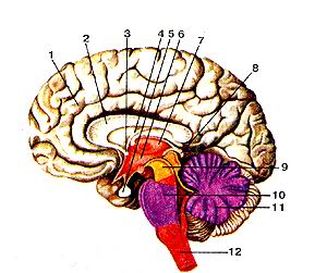
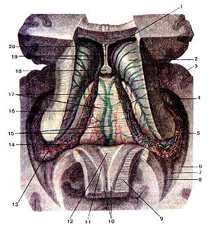
Small. 253. Brain (vertical growth):
1 - corpus callosum; 2 - star; 3 - thalamus; 4 - dah of the middle brain; 5 - nipple-like body; 6 - water supply to the middle brain;
7 - nіzhka to the brain; 8 - zorove overbaptism; 9 - IV crotch; 10 - hipophys; 11 - mest; 12 - cerebellum
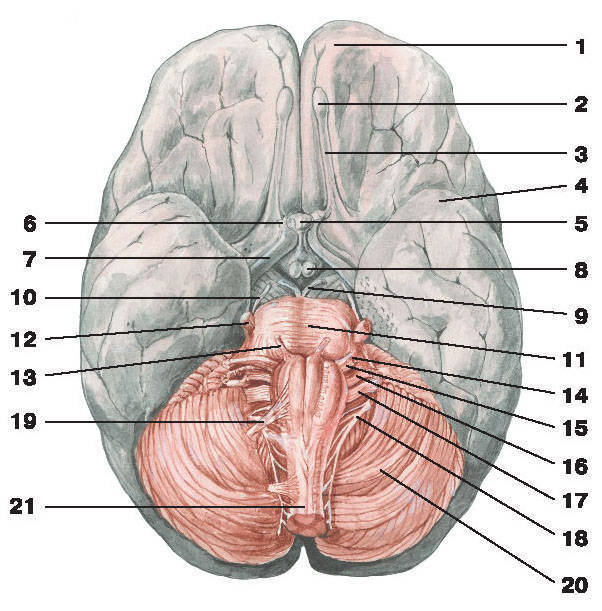
Small. 254. Brain (bottom view):
1 - frontal part; 2 - snuff cybulin; 3 - scent tract; 4 - skroneva part; 5 - hipophys; 6 - healthy nerve;
7 - healthy path; 8 - nipple-like body; 9 - gory nerve; 10 - block nerve; 11 - mest; 12 - tripartite nerve;
13 - a nerve to introduce; 14 - facial nerve; 15 - frontal-ultkovy nerve; 16 - the ulopharyngeal nerve; 17 - bloody nerve;
18 - dodatkovy nerve; 19 - pidyazic nerve; 20 - cerebellum; 21 - dovgastiy brain
Small. 255. Brain (transverse growth):
1 - ost_vets; 2 - shkaralupa; 3 - fence; 4 - zvnishnya capsule; 5 - blidiy kulu; 6 - III crotch;
7 - chervone core; 8 - pokrishka; 9 - water supply to the middle brain; 10 - dah middle mozu; 11 - hipocampus; 12 - cerebellum
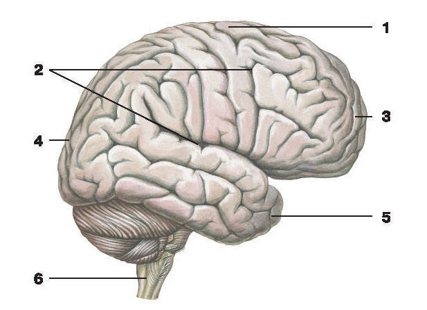
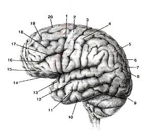
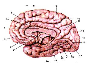
Small. 258. Parts of the brain (side view):
1 - tim'yana part; 2 - furrows of the brain; 3 - frontal part; 4 - part of the city;
5 - skroneva part; 6 - spinal cord
Small. 260. Mozok (side view):
1 - nіzhka to the brain; 2 - the upper surface of the pivucleus of the moss; 3 - hipophiz; 4 - bili plates; 5 - mest; 6 - toothed core;
7 - bila speech; 8 - dovgastia brain; 9 - olive kernel; 10 - the lower surface of the pivucleus of the moss; 11 - spinal cord
Small. 262. For the brain:
1 - upper lobe of the corn; 2 - pіramіdny tract; 3 - nіzhka kіntsevoy marz; 4 - middle nizhka of the corn; 5 - mest;
6 - lower lobe of the corn; 7 - olive; 8 - pyramida; 9 - front middle slit

Wonder so much:
Nervous system
Central nervous system Spinal cord
dimpled brain
Parts of pivkul great mozka
Prodigal brain
misst
lobe
IV crotch
middle brain
middle brain
The conduits of the nervous system
Shells and between shells
Peripheral nervous system
cranial nerves
Spinal cord nerves
Vegetative nervous system
The cerebrum (encephalon) (Fig. 258) will grow in the empty cerebral skull. The middle wall of the brain of a grown-up human becomes approximately 1350. The wine has an ovoid shape through the protruding frontal and longitudinal poles.
On the outer lump of the upper lateral surface of the brain (facies superolateralis cerebri), the number and size of the furrow (sulci cerebri) grow in number and form (Fig. 258). Above, do not go into them, there is a pavutin of the sheath of the brain. From the poles of the pole to pass the transverse slit of the great brain, before which is the bed of the brain, which is the most important center for the coordination of the arms. For the middle line, the brain undergoes a late fissure (fissura longitudinalis cerebri), which is distributed on the right and left of the brain (hemispherium cerebri dextrum et sinistrum). The lower surface (fasies inferior cerebri) is characterized by a folding top.
At the empty skull, the spinal cord is trivialized with a gastimose, which can be used to take revenge on the vertebral column and dical center. The higher growths in the lower part of the brain and the cerebellum are joined one by one behind the auxiliary bridge, which is removed from the large brain. The lobe grows backwards out of the cichs. From the front edge of the bridge forward and towards the side of the brain to enter the brain (pedunculis cerebri) (Fig. 253, 255, 260, 262), to encircle the middle fossa. In front of the fossa, the nipple-like til (corpus mamillare) (fig. 253, 254) grow out, which represents the swelling of the day and is presented to the scent analyzer. In front of the papillary bodies, there is a gray hump (tuber cinereum), until the lower cerebral appendage creeps behind the auxiliary funnel, the titles of hypophysis (Fig. 253, 254, 260) and a neuroendocrine organ. 12 pairs of cranial nerves, rosted on the lower surface of the brain, are carried to the peripheral nervous system.
Empty the brain, which is a surplus of cerebral microorganisms, which is formed in the membrane period, and is stored in the brain. Dovgastia brain, posterior cerebrum, which includes the cerebellum and myst, grows in a single empty space, called the IV lug (Fig. 253). The emptying of the middle brain is called the aqueduct of the middle brain (aquaeductus mesencephali). Before her, the middle brains grow out, and above it there are guys humps, which make a quadruple. Porozhnin of the intermediate brain is called the III slurry and includes the thalamus, neuroendocrine organisms (hypophyses with rostasis between the upper burrows) and deyakі іnshі structures. Kintseviy moss stores the great puffs of the brain, separated by adhesions, most of the corpuscles tilo. At the tovschі pіvkul, they fill up the little shlunochki.
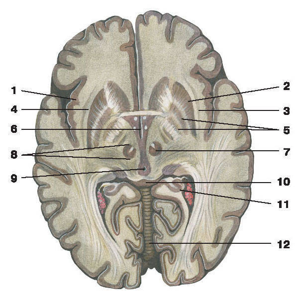
Small. 338. Great brain (cerebrum). The projection of bichny shlunochkiv on the surface of the great pivkul
brain. Top view. I-lobova part; 2-center boron; Th-b-horse slunk; 4-for-tilochnaya part; 5-back-n_y rіg of the creepy duck; 6-IV crotch; 7-water supply to the brain; 8-III croaker; 9-central part of the b_chny shlunochka; 10-lower ryg of the bicorny clown; 11-front rig of the crawler.
Fig. 338. Cerebrum. The projection of bichons on the surface of the great brain. Top view. I-lobus fronlalis; 2-sulcus centralis; 3-ventriculus lateralis; 4-lobus occip-italis; 5-cornu posterius ventriculi lateralis; 6-IVventriculus; 7-aguaecluc-tuscerebri; 8-111 ventriculus; 9-pars centralis ventriculi lateralis; 10-cornu inferius ventriculi lateralis; 11-cornu anterius ventriculi lateralis.
Fig. 338. Cerebrum. Projections of lateral ventricles on the surface of
the hemispheres of the cerebrum. Superior aspect. I-frontal lobe; 2-ccntral fissure; 3-latcral ventricle; 4-occipital lobe; 5-back horn of the lateral ventricle; 6-IV ventricle; 7-aqucduct of brain; 8-111 ventricle; 9-central part of lateral ventricle; 10-inferior horn of lateral ventricle; 11 -anterior horn oflateral ventricle.
Small. 339. Brain (cerebrum). Sagittal development. View from the medial side.
I-drink of the great brain; 2-corpus callosum; 3-front (bila) spike; 4-star brain; 5-hypophyse; 6-empty midbrain (III corkscrew); 7-thalamus; 8-ergyphys to the brain; 9-middle brain; 10th place; 1 (-cerebellum; 12-dovastia cerebrum.
Fig. 339. Cerebrum.
Sagittal development. View from the medial side, l-hemispherium ccrebri: 2-corpus callosum; 3-comissura (alba) anterior; 4-fornix medullae spinalis; 5-hypophysis; 6-cavum encephali intermedii (3 ventriculus); 7-thalamus; 8-epiphysis encephali; 9-medulla media; 10-pons: 11-cerebellum; 12-medulla oblongata.
Fig. 339. Cerebrum. Sagittal section. From the medial side. 1-cerebral hemisphere; 2-corpus callosum; 3-anterior (white (commissure; 4-fornic; 5-hypophysis; 6-space of diencephalon (111 ventricle); 7-diencephalon: 8-pineal body of brain; 9-midbrain; 10-pons; 1 l-cere- bellum; 12-medulla oblongata.
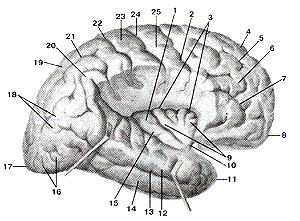
Small. 340. Upper-lateral surface of the great
brain.
I-precentral beard; 2-precentral zvivina 3-central beard; 4-postcentral zivina; 5-top; tim'yana part; 6-internal borozy; 7-lower tim'yan * part; 8-kutova zvivina; 9-pole pole; 10-lower skroneva zvivin; 11-lower skroneva beard; 12-middle skronev zvivin; 13-top skroneva zvivin; 14-lateral beard; 15-orbital part; 16-lower lobova zvivin; 17-lower lobova beard; 18-middle lobova zvivin; 19-upper lobova beard; 20-top lobova zvivina ..
Fig. 340. Upper-lateral surface of the great
brain.
1-sulcus precentralis; 2-gyrus precentralis; 3-sulcus centralis; 4-gyais postcentralis; 5-lobulus parietalis inferior; 6-sulcus interparietalis; 7-lobulus parietalis inferior; 8-gyrus angularis; 9-polus occipitalis; 10-gyrus temporalis inferior; 11-sulcus temporalis inferior; 12-gyrus temporalis medialis; 13-gyrus temporalis superior; 14-sulcus lateralis; 15-pars orbitalis; 16-gyrus frontalis inferior; 17-sulcus frontalis inferior; 18-gyrus frontalis medialis; I9-sulcus frontalis superior; 20-gyrus frontalis superior.
Fig. 340. Superiolateral surface of the hemisphere of the cerebrum. 1-precentral sulcus; 2-precentral gyrus; 3-central sulcus; 4-postcentral sulcus; 5-upper parietal lobule; 6 interparietal sulcus; 7-lower parietal lobule; 8-angular gyrus; 9-occipital pole; 10-inferior temporal gyrus; ll-inferior temporal sulcus; 12-middle temporal gyrus; 13-superior temporal gyrus; 14-lateral sulcus 15-orbital part; 16-inferior frontal gyrus; 17-inferior frontal sulcus; 18-middle frontal gyrus; 19-superior frontal sulcus; 20-superior frontal gyrus.
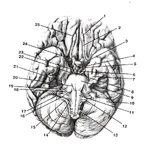
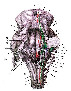
Small. 341. The lower surface (base) of the brain
Mystery to the origin of the cranial nerves. I-snuff cybulin; 2-scent tract; 3-front (perforated speech; 4-gray hillock; 5-healthy tract of 6-nipple-like til; 7-three-part vuzol; 8-back perforations; 9-mest; 10-cornsus; 11-pyramida 13-; 12-olive spinal nerve; 14th pidyom) ichny nerve; 15-dodatkovy? nerve; 16-vagus nerve; 17th ipsiopharyngeal nerve; 18-before the virino-ultkovy nerve; 19 facial nerve; 20-part nerve; 21-part nerve; 22-block nerve; 23-okoruhovy (nerve; 24-healthy nerve; 25-nyukhova boroznaya
Fig. 341. The lower surface (base) of the brain
miscarriage of cranial nerve roots, l-bulbus olfactorius; 2-tractus olfactorius; 3-substantia pertbrala anterior; 4-tubercinercum; 5-tractusopticus; 6-sofa mamillare; 7-gan-glion trigeminale; 8-substantia perforate posterior; 9-pons; 10-cerebcl-lum; ll-pyramis; 12-oliva; 13-nervus spinalis; 14-nervus hypoglossus; 15-nervus accessorius; 16-nervus vagus; 17-nervus glossopharyngeus; 18-nervus vestibulocochlearis; 19-nervus facialis; 20-nervus abduccns; 2 l-nervus trigeminus; 22-nervus trochlearis; 23-nervus oculomotorius; 24-nervus opticus; 25-sulcus olfactorius.
Fig. 341. Lower surface of the cerebrum with the origins of the cranial
nerves.
I-olfactory bulb; 2-olfactory tract; 3-anterior perforated substance; 4-tuber cinerum; 5-optic tract; 6 mammillary bodies; 7-trigeminal ganglion; 8-posterior perforated substance; 9-pons; 10-cerebellum; II-pyramid; 12-olive; 13-spinal nerve; 14-hypoglossal nerve; 15-accessoriusnerve; 16-vagus; 17-glossopharyngeal nerve; 18-vestibu-locochlear nerve; 19-facial nerve; 20-abduccnt nerve; 21 trigeminus nerve; 22-troclear nerve; 23-oculo-motor nerve; 24-optic nerve; 25-olfactory sulcus.
Small. 342. Media and lower surfaces
great brain.
1-star; 2-dzob of the corpus callosum; 3-colony corpus callosum; 4-stovbur of corpus callosum; 5-borne corns; 6-belt zivina; 7-upper frontal zvivin; 8-sub-parietal boron; 9-paracentral part; 10-belt bearded; P-pre-wedge; 12-tim'yano-potilichna beard; 13-wedge; 14-spur boron; 15-movny zvivina; 16-medіyna tilichno-skroneva zvivin; 17-sided-skroneva beard; 18-lateral zylochno-skroneva zvivina; 19-boron hipocampus; 20-paragіppo-campal zivina.
Fig. 342. Media and lower surfaces
great brain.
l-fornix; 2-rostrum sofopv callosi; 3-genu sofopia callosi; 4-truncus sofopv callosi; 5-sulcus sophops callosi; 6-gyrus cingulis; 7-gyrus fronlalis superior; 8-sulcus cingulis; 9-lobulus paracentralis; 10-sulcus subparietalis; 11-precuneus; 12-sulcus occipitoparietalis; 13-cuneus; 14-sulcus calcarinus; 15-gyrus lingvalis; 16-gyrus occipitotemporalis mcdialis; 17-sulcus occipitotemporalis; 18-gyrus occipitotemporalis lateralis; 19-sulcus hippocampi; 20-gyrus parahippocampalis.
Fig. 342. Medial and lower surfaces of the hemisphere of the cerebrum.
l-fornix; 2-rostrum (ofsofiz callosum); 3-genu (ofsofіvcallosum); 4-trunk; 5-sulcus of sofiv callosum; 6-singulate gyrus; 7-superior frontal gyrus; 8-singulate sulcus; 9-paracentral lobule; 10-subparielal sulcus; 11-medial occipitotemporal gyrus; 12-parieto-occipital sulcus; 13-cuneus; 14-calcarine sulcus; 15-gyrus; ! 6-medial occipitotemporal gyrus; 17-occipitotemporal sulcus; 18-lateral occipitotemporal gyrus; 19-hippocampal suicus; 20-parahippicampal gyrus.
Small. 343. Ostrіvets (insula). Ostrіvkova part. View from the lateral side. Part of the tim'yano and frontal part
you have seen. The skrone part is pulled down to the bottom.
1-island; 2-precentral boron; 3-circle boroznaya island; 4-upper frontal zvivin; 5-upper lobova beard; 6-middle lobova zvivin; 7-lower lobova beard; 8-front (front) pole; 9-short zivini island; 10-time island; 11-pin pole; 12-top skroneva zvivin; 13-upper skroneva beard; 14-middle skronev zvivin; 15-dovga zvivina island; 16-lateral potilichny zivini; 17-sided (back) pole; 18-kutova zvivina; 19-upper tim'yan part; 20-supra-marginal zivin; 21-intra-parietal boron; 22-post-central boron; 23-possentralny zvivina; 24-center boron; 25-presetting zivine.
Fig. 343. Ostrіvets. Ostrіvkova part. View from the lateral side. The part of the temporal and frontal part is visible. Skronev's part otgyanuga to the bottom.
1-insula; 2-sulcus precentralis; 3-sulcus orbicularis insulae; 4-gyrus Ironialis superior; 5-sulcus I "rontalis superior; 6-gyrus trontalis medi-alus; 7-sulcus I" rontalis inferior; 8-polus fromalis (anterior); 9-gyri breves insulae; 10-limen insulae; 11-polus temporalis; 12-gyrus temporalis superior; 13-sulcus temporalis superior; 14-gyrus temporalis medi-alus; 15-gyrus longus insulae; 16-gyri occipilales laterales; 17-polus oceipilalis (posterior); 18-gyrus angylaris; 19-lobulus parictalis superior; 20-gyrus supramarginalis; 21-suleus intraparietalis; 22-sulcus post-eentralis; 23-gyrus postcenlralis; 24-sulcus ccnlralis; 25-gyrus precentralis.
Fig. 343. Insula. Insular lobe. Lateral aspect. Part of the parietal and frontal lobes are removed. Temporal lobe
replaced downstaires.
l-insula; 2-preeentral sulcus: 3-cireular sulcus of insula: 4-superior frontal gyms; 5-superior frontal sulcus; 6-middle frontal gyrus; 7-infe-rior frontal sulcus; 8-frontal (anterior) pole; 9-short gyrus of insula; IO-limen insulae (insular threshold); 11-temporal pole; 12-superior temporal pole; 13-superior temporal sulcus; 14-middle temporal gyrus; 15-long gyrus of insula; 16-latcral occipital gyri; 17-occipilal (posterior) pole; 18-angular gyrus; 19-superior temporal lobule; 20-supramarginal gyrus; 21 intraparietal gyrus; 22-postcentral sulcus; 23-postcentral gyrus; 24-central gyrus; 25-precentral gyms.
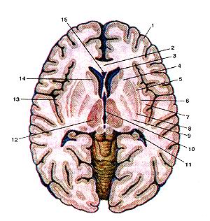
Small. 344. Basal (subcortical) vuzli (nuclei basales) and inner capsule (capsula interna) on a horizontal opening
brain. Top view
1-bark of the great mozu (cloak); 2-colony corpus callosum; 3-front rig of the bicorny clown; 4-inner capsule; 5-caruzh-va capsule; 6-ofada; 7th capsule itself; 8-shkaralupa; 9-blade ball; IO-III slut; II-rear rig of the bicorny clown; 12-thalamus (green hillock); 13-kirkova river (bark) island; 14-head of the caudate nucleus; 15-empty spacer.
Fig. 344. Basal nuclei and internal capsule on the horizontal growth of the brain. Top view. I-cortex cerebri; 2-genu corporis callosi; 3-cornu anterius ventriculi lateralis; 4-capsula inlerna; 5-capsula extema; 6-claustnim; 7-capsula externa; 8-pulamen; 9-globus pallidus; 10-venlriculustetrius; 11-comu posierius ventriculi lateralis; 12-thalamus; 13-substantiacorticalis (cortex) insulae; 14-caput-nuclei caudatae; 15-cavum septi pellucidi.
Fig. 344. Basal (infracortical) ganglions with internal capsule on the
cross-section of the cerebrum. Superior aspect. 1-cerebral cortex; 2-genu of callous body; 3-anterior horn of lateral ventricle; 4-internal capsule; 5-external capsule; 6-claustrum; 7-extreme capsule; 8-putamen; 9-globus pallidus; 10-111 ventricle; II-posterior horn of lateral ventricle; 12-thalamus; 13-insular cortex; 14-head (of caudate nucleus); 15-cave (of septum pellucidum).
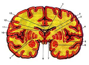 Small. 345. Basalis (pіdkіrkovі) umi (nuclei basales) on the frontal development of the brain, development of fractures on івні
nipple bodies.
1-Sudinne gossip of a bicorny louse (central part); 2-thalamus; 3-internal capsule; 4-bark island; 5-ofada; b-blinkingly; 7-healthy pass; 8-nipple-type tilo; 9-blade ball; 10-shkaralupa; 11-star to the brain; 12-tailed nucleus; 13-callus tilo.
Fig. 345. Basal nuclei on the frontal height of the head
to the brain, the growth of fractures on the pits of the mastoid bodies. l-plexus choroideus ventriculi laleralis (pars cenlralis); 2-thalamus; 3-capsula inlerna; 4-cortex insulae; 5-clauslrum; 6-corpus amyg-daloidcum; 7-lraclus opticus; 8-corpus mammillare; 9-globus pallidus; 10-putamen; 11-fornixcerebri; 12-nucleuscaudatus; 13-corpuscallo-
Fig. 345. Basal ganglions on the front-section ot the cerebrum. The
discission is made on the level ot "mammilary bodies, l-choroid plexus of lateral ventricle (central pan); 2-thalamus; 3-inter-nal capsule; 4-insular cortex, 5-claustruin; 6-amygdaloid body; 7-opli -cal tract; 8-mammilary body; 9 -globus pallidus; 10-putamen; II-fornix; 12-tail (of caudate nucleus); 13-corpus callosum.
Small. 346. Ventriculi laterales and Sudinna
the basis of the third louse (tela chorioidea ventriculi tertii). view
above. The corns is thin and thin to the brain.
opened in
I-front rig of the bicorny duck; 2-tailed nucleus; 3-sided gossip in the central part of the right side duck; 4-nіzhki hіpocampus; 5-sudinne gossip in the lower pink bicon; 6-collateral p_densennya; 7-bird spur; 8-cybulin of the dorsal horn; 9-corpus callosum; 10-tilo star; 11-nіzhka zvod; 12-spike to the star; 13-villous artery; 14-great mozkov vein; 15-internal mozkov vein; 16-upper thalamos griarny vein; 17-vessel base of the third lander; 18-stop to the star; 19-plate with a septum gap; 20-empty space for the septum.
Fig. 346. Biches of a dugout and a ship's base of the third dugout. Top view. Calluses thither and thither to the brain
re-registration and entry into the list.
l-cornu anterius venlriculi lateralis; 2-nucleus caudatus; 3-choroideus ventriculi (partis centralis); 4-pes hippocampi; 5-plexus choroideus ventriculi lateralis (cornu interior); 6-eminentia collaterals; 7-calcar avis; 8-bulbus cornus posterioris; 9-corpus callosum; 10-corpus forni-cis; 11-crus fornicis; 12-comissura fomicis; 13-arteria choroidea; 14-vena cerebri magna; 15-vena cerebri interna; 16-vena thalamostriara superior; 17-tela vascularis ventriculi tertii; 18-columnae tornicis; 19-lamina septi pellucidi; 20-cavum septi pellucidi.
Fig. 346. Lateral ventricles and choroid tela of the third ventricle. Superior aspect. The corpus callosum and body of fornix are incised
and put to backward.
l-antcroir horn of lateral ventricle; 2-caudate nucleus; 3-choroid plexus within the central part of the right lateral ventricle; 4-pes of hippocampus; 5-choroid plexus within the lower horn of the lateral ventricle; 6-col-lateral eminence; 7-calcurine spur; 8-bulb of occipital horn; 9-corpus
callosum; lO-body (offornix); 11-cms (of fbrnix); 13-choroid artery; 14 great cerebral vein; 15-internal cerebral vein; 16-superior talamostriate vein; 17-choroid tela of the third ventricle; 18-columns (oftornix); 19-lamina (of septum pellucidum); 20-cave (ot "septum pelluciduni).
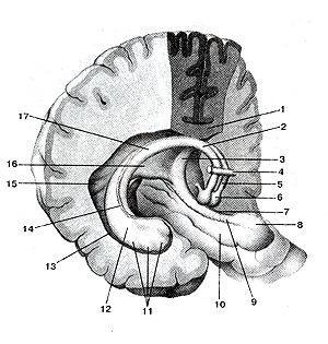
Small. 347. Fornix and hippocampus.
Bil above and three to the side.
1-wash tilo; 2-tilo zvod; 3-nіzhka zvod; 4-anterior commissure; 5-stovp zvod; b-SOSyevidshk tilo; 7-fringe Gippo kampa; 8-hook; 9-tooth Zivin; 10-naragishyukammalna zivina; 11-nіzhka hіpocampus; 12-hipocamp; 13-Bokono and pipes (rozkritiy); 14-fringe of the hipocampus; 15-piece spur; 16-spike to the zvod; 17-nіzhka zvod.
Fig. 347. Star and hipocampus. Zgori and Trocha view from the side. 1-corpus callosum; 2-corpus fornicis; 3-crus tbrnicis; 4-commissur< anterior; 5-collumna fornicis;
6-corpus mammillare; 7-fimhria hip pocampi; 8-uncus; 9-gyrus denial us; I0-gyrusparahippocampialis;
i I-crus hippocampi; 12-hippocampus; 13-ventriculus lateralis (вскрыт): 14-fhnbria
hippocampi; 15-calcaravis; 16-comissura fornicis; 17-crus lornicis.
Fig. 347. The lornix and the hippocampus. Supero-laleral aspect. 1-corpus callosum; 2-body (of fornix); 3-crus (of fornix); 4-anterior commissure; 5-columns (of fornix); 6-niammilary body; 7-llmbria (of hippocampus); 8-uncus; 9-dental gyrus; 10-parahippocampal gyms; ll-pes (of hippocampus); 12-hippocampus; 13-lateral ventricule (opened); 14-llmbria (of hippocampus); 15-calcurine spur; 16-com-missure (of fornix); 17-pes (of fornix).
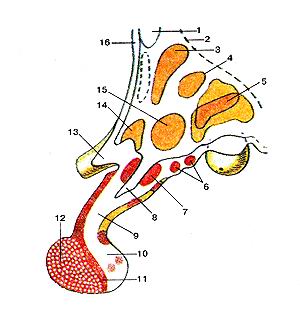
Small. 348. Hypothalamus (hypothalamus) and hypophysis
(Hypophisis) on sagittal growth. The nucleus of the hypothalamus. 1-anterior commissure; 2-hypothalamic beard; 3-peri-ventricular nucleus; 4-upper medial nucleus; 5-posterior nucleus; 6-siro-tubercle of the nucleus; 7-funnel core; 8-sinking funnel; 9-crow-ka hypophysis; 10-posterior part of the hypophysis; 1 [-intermediate part of the hypophysis; 12-front part of the hypophysis; 13-zorove overbaptism; 14-supervisory nucleus (supraoptic); 15-anterior hypothalamic nucleus; 16-terminal plate.
Fig. 348. Hypothalamus and hypophysis on sagittal growth.
The nucleus of the hypothalamus.
l-commissura anterior; 2-sulcus hypothalamicus; 3-nucleus paraven-iricularis; 4-nucleus medialis superior; 5-nucleus postrior; 6-nuclei tuberalis; 7-nucleus infundibularis; 8-recessus untundibuli; 9-unfundibulum; 10-lobus posterior (hypophisis); 11-pars intermedia (hypophisis); 12-lobus anterior (hypophisis); 13-chiasma optica; 14-nucleus supraorbitalis; 15-nucleus anterior hypothalami; 16-lamina terminalis.
Fig. 348. Hypothalamus and hypophysis (pituatry gland).
Nuclei of hipotalamus. Sagittal section
1-anterior commissure; 2-hypothalamic sulcus; 3-paraventricular nucleus; 4-superomedial nucleus; 5-postrior nucleus; 6-tuberal nucleus; 7-infundibular recess; 8-infundibular (arcuate nucleus); 9-infundibulum; l () - postrior lobe (of hypophysis); 11-pars intermedia (of hypophysis); 12-anterior lobe (of hypophysis); 13-optic chiasm; 14-supra-optic nucleus; 15-anterior hypothalamic nucleus; 16-terniinal lamina.
Small. 349. Middle MO3r (mesencephalon). Cross rosrіz. 1-dah middle brain; 2-pokrishka middle moss; 3-base-nya nіzhki to the brain; 4-chervone core; 5-chorna speech; 6-core of the okoruha nerve; 7-dodatkovo nucleus of the okoruha nerve; 8-cross pokrishki; 9-gory nerve; [0-Lobno-Mostovoi way; 11-core-core way; 12-cork-sp_shumo zgovy pug; 13-sided-skronevo-tim'yano-bridge-voyi; 14-med_yna loop; 15-handle of the lower hump; 16-nucleus of the spinal cord of the tripartite nerve; 17-upper hump; 18-water supply to the middle brain; 19-central syra speech.
Fig. 349. My middle Cross-section 1-tectum mesencephalicum; 2-tegmenlum mesencephalicum; 3-basis pedunculi celebri; 4-nucleus ruber; 5-subslanlia nigra; 6-nucleus nervi oculornotprii; 7-nucleus oculomotorium accessorius; 8-decussaliones tegmenti; 9-nervus oculomolorius; 10-traclus fronlopontinus; 11-trac-tuscorticonuclearis; 12-lracluscorticospinalis (pyramidalis); 13lraclus occipilolemporoparietoponlinus; 14-lemniscus medialis; 15-brachium colliculi inferioris; 16-nucleus Iractus mesencephalici nervi trigemi-nalis; 17-colliculus cranialis (superior); 18-aqueductus mesencephali (celebri); 19-subslanlia grisea ceniralis.
Fig. 349. Midbrain. Transversal section.
1-lectal pluie; 2-tegmenlum ofbrain; 3-base of peduncle (of brain); 4-red nucleus; 5-substantia nigra; 6-oculomotor nucleus; 7-accessory oculomotor; 8-decussation tegmenlalis; 9-oculomolor nerve; 10-fron-topontine tract; 11-corticonuclear tract; 12-conicospinal tract; 13-occipilotemporoparietal tract; 14-medial lemniscus; 15-brachium of interior colliculus; 16-spinal nucleus of trigeminal nerve; 17-superior colliculus; 18-aqueduct ofbrain (midbrain); 19-substanlia grisea (cen-Iral graysubslancc).
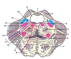
Fig. 350. MocT (pons).
Lateral growth on the edge of the upper cerebral vitril. 1-upper cerebral sail; 2-upper lobe; 3-posterior lateral bundle; 4-central pokrishechno way; 5-lateral loop; 6-med_yna loop; 7-way bridge fibers (first way); 8-part nerve; 9-nucleus of the facial nerve; 10-core of the external nerve; II-facial nerve; 12-triple nerve; 13-rokova nucleus of the tricular nerve; 14-upper salivary nucleus; 15-bridge nucleus of the tricyclic nerve; 16-core single way; 17-IV croaker.
Fig. 350. Міст.
Lateral growth on the level of the upper cerebral vitril. 1-velum medullare rostralis (superius); 2-pedunculus cerebellaris superior; 3-fasciculus longitudinalis posterior; 4-tractus tegmentalis cen-tralis: 5-lemniscus lateralis; 6-lemniscus medialis; 7- (tractus pyrami-dalis) librae longitudinales pontis; 8-nervus abducens; 9-nuclcus nervi lacialis; 10-nucleus nervi abducentis; ll-nervus facialis; 12-nervus trigeminus; 13-nucleus motorius nervi trigemini; 14-nucleus salivato-rius; 15-nucleus pontinus nervi trigemini; 16-nucleus solitarius; 17-IV ventriculus.
Fig. 350. Pons.
Transversal section at a level of the upper cerebral cloth. 1-superior medullary velum; 2-superiorcerebellar peduncle; 3-posteri-or longitudinal fascicle; 4-central tegmental tract; 5-lateral Icmniscus; 6-medial lemniscus; 7-pyramidal tract; 8-abducent nerve; 9-nucleusof facial nerve; 10-nucleus of abducent nerve; 1 l-facial nerve; 12-trigem-inal nerve; 13-nucleus of trigeminal nerve; 14-superior salivatory nucleus; 15-nucleus pontine of trigeminal nerve; 16-solitary nucleus; 17-IV ventricle.

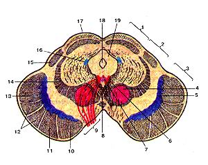
Small. 351. My Dovgasti (medulla oblongata).
Porechny Razrіz on Rivnі olives.
I-quarter crotch; 2-dorsal nucleus of the blukus nerve; 3-core of the vestibular nerve; 4-core of a single path; 5-posterior (dorsal) spinal cord; 6-spinal nucleus of the tricyclic nerve; 7-spinal cord path of the triple nerve; 8-nucleus of the pid'yazic nerve; 9-Olive core; 10th hour; 11-cortical-SPINAL MUSCULAR pug (pyramid); 12-med_yna loop; 13-PIDYASIC nerve; 14-front outer arc fibers; 15 sub-core; 16-dorsal-thalamic and dorsal-on-kryshkovy way; 17-bloody nerve; 18-ventralpium (anterior) spinal cord path.
Fig. 351. Dovgasti mozok. Porechny Razrіz on Rivnі olives. 1-IV ventriculus; 2-nucleus dorsalis nervi vagii; 3-nucleus vcstibularis; 4-nucleus solitarius; 5-traetus spinocerehellaris posterior (dorsalis); 6-nucleus spinalis nervi trigemini; 7-tractus spinalis nervi trigemini; 8-nucleus nervi hypoglossi; 9-nucleus olivaris; 10-oliva; 11-tractus corti-cospinalis; 12-lemniscus medialis; 13-nervus hypoglossus; 14-llbrae arcuatae externae anteriores; 15-nucleus atnbiquus; 16-tractus spinothalamicus et spinotectalis; 17-nervus vagus; 18-tractus spin-occrebellaris ventralis (anterior).
Fig. 351. Myelencephalon. Transverse section at a level of the olives. 1-fonh ventricle; 2-dorsal nucleus of vagus nerve; 3-nucleus of vestibular nerve (vestibular nucleus); 4-solitary nucleus; 5-posterior dor-socerebellar tract; 6-spinal nucleus of trigeminal nerve; 7-spinal tract of trigeminal nerve; 8-nucleus of hypoglossal nerve; 9-olivary; 10-olive; 1 l-corticospinal tract; 12-medial lemniscus; 13-hypoglossal nerve; 14-anterior external arcuate fibers; 15-nucleus ambiquus; 16-spinothalam-ic and spinotectal tracts; 17-vagus nerve; 18-ventral spinocerebellar tract.
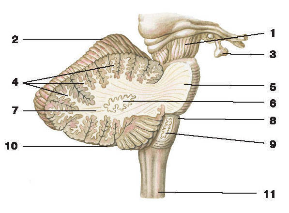
Small. 352. Crab (cerebellum). Top view. 1-worm of a corn; 2-nodule of the corn; 3-lines (furrows) of the corn; 4-leaf corns; 5-horizontal shelf; 6-posterior virіzka moss; 7-bottom half-part; 8-top nap_vm_shayachna part; 9-chotirikutna patch; 10-lower humps dahu middle brain; 11-upper hump; 12-epiphiz; (3-tala-musi; 14th third lingo.
Fig. 352. Mozok. Top view.
1-vermis cerebelli; 2-hemispherium cerebelli; 3-fissurae cerebelli; 4-folia cerebelli; 5-fissura horizontalis; 6 incisura cerebelli posterior; 7-lobulus semilunaris inferior; 8-lobulus semilunaris superior; 9-lobulus quadrangularis; 10-coliculus interior (menescephalici); 11-coliculis superior; I2-epiphysis; 13-thalami; 14-ventriculus tertius.
Fig. 352. Cerebellum. Superior aspect.
I-vermis of cerebellum; 2-hemisphere of cerebellum; 3-cerebellar fissures; 4-folia of cerebellum; 5-horizontal fissure; 6-posterior incisure of cerebellum; 7-inferior semilunar lobule; 8-superior semilunar lobule; 9-quadrangular lobule; 10-inferior colliculus of midbrain; II-superior colliculus (of midbrain); 12-pineal body; 13-thalamuses; 14-third ventricle.
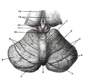
Small. 353. Cerebellum, mesencephalon
and intermediate brain (diencephalon). Top view. pіvkulі
the great brain is seen. The lobe of roscritium is horizontal
rozrіzom, we will carry out on a level horizontal slit
corns.
1-cerebellar-red-nuclear path; 2-core of the tent; 3-worm (corn); 4-kulyaste core; 5-corky core; 6-mozcova t "pro (moss); 7-gate of the toothed nucleus; 8-bile plates; 9-tooth core; 10-upper lobe; 11-lump of the upper cerebral vitril; 12-lower hump (middle brain); 13-upper hump; 14-epіphіz to the brain (pineal tіlo); 15-tricycle of the age; 16-thalamus; 17th third slunker.
Fig. 353. Mozok, middle brain and intermediate brain. view
above. The inspiration of the great brain is visible. rozcritium lobe
horizontal growth, carried out on the
the corns went horizontally.
l-tractus cerebellorubralis; 2-nucleus lasligi; 3-vermis cerebelli; 4-nucleus globosus; 5-nucleus emboliformis; 6-corpus medutlare; 7-hilum nuclei dentati; 8-laminae albae; 9-nucleus dentatus; 10-pedun-culus cerebellaris superior; ll-frenulum veli medullaris rostralis (superioris); 12-colliculum inferior (mesencephali); 13-colliculum superior; 14-sof_5 pineale; 15 trigonum habenulare; 16-thalamus; 17-ventriculus tertius.
Fig. 353. Cerebellum, medial brain and the betweenbrain. Upper sight. Hemispheres of the cerebrum are cut of. Cerebellum "s anatomized with horizontal cut; performed at a level of the horizontal fissure of the cerebellum.
1-cerebellorubral tract; 2-fastigal nucleus; 3-vermis of cerebellum; 4-globus nucleus; 5-emboliform nucleus; 6-medullary body (of cerebellum); 7-hilum of dentale nucleus; 8-white substans of cerebellum; 9-den-tale nucleus; 10-superior cerebellar peduncle; ll-frenulum of superior medullary vellum; 12-inferiorcolliculus; 13-superiorcolliculus; L4-pineal body of brain; 15-habenulartrigone; 16-thalamus; 17-third ventricle.
Small. 354. Quarter crotch (venticulusquartis) is the base of the fourth crotch (tela chorioidea ventriculi quarti).
Top view.
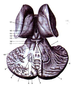
1 uvicle of the corn; 2-srhn_y brain sail; 3-quarter crotch; 4-middle nіzhka of the corn; 5-Sudinne gossip of the fourth crook; 6-hump of wedge-like core; 7-tubercle thin nucleus; 8-rear prom_zhna borozna; 9-wedge-shaped beam; 10-point (lateral) cord; 11-thin beam; 12-rear middle bordered; 13-posterior lateral furrowed; 14-middle hole (aperture) of the fourth aperture; 15-vascular base of the fourth crotch; 16-upper (front) lobe of the corn; 17-block nerve; 18-lower hump (dahu middle brain); 19-lump of the upper cerebral vitril; 20-top hump (dahu middle moss).
Fig. 354. Quarter lander and fourth base
slunk. Top view.
l-lingula cerebelli; 2-veilum medullare rostralis; 3-IV ventriculus; 4-pedunculus cerebellaris medius; 5-plexus choroideus ventriculi guarti; 6-tuberculum cuneatum; 7-tuberculum gracile; 8-sulcus intermedius posterior; 9-fasciculus cuneatus; 10-fasciculus lateralis; 11-fasciculus gracilis; 12-sulcus meduanus posterior; I3-sulcus dorsolateralis; 14-foramen (apertura) mediana ventriculi guarti; 15-tela choroidea ventriculi guani; 16-pedunculus cerebellaris rostralis (superior); 17-nervus trochlearis; 18-colliculus inferior (tecti menescephali); 19-frenulumveli medullaris rostralis; 20-colliculus superior (tecti menescephali).
Fig. 354. The fourth ventricle and tela choroidea
of the fourth ventricle. Superior aspect.
l-lingula; 2-superior medullary velum; 3-fourth ventricle; 4-middle cerebellai peduncle; 5-choroid plexus of fourth ventricle; 6-tubercle of cuneale nucleus; 7-tubercle of gracile nucleus; 8-posterior intermediate sulcus; 9-cuneate fasciculus; 10-lateral funiculus; 11-gracile fasciculus; 12-posterior median sulcus; 13-posterolateral sulcus; 14-median aperture; 15-tela choroidea of the fourth ventricle; 16-superior (anterior) cerebellar peduncle; 17-trochlear nerve; 18-inferior colliculus; 19-frenulum of superior medullary vellum.
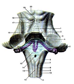
Small. 355. Rhomboid fossa (fossa rhomboidea). The posterior surface of the bridge and the brain, projection of the nuclei (s)
cranial nerves to a rhomboid fossa.
1-dodatkovo (parasympathetic) nucleus (i.) Of the okorukhovy nerve; 2nd. gory nerve; 3rd. block nerve; 4th. midbrain way of the threefold norn; 5-two-fold "tricular nerve; 6-bridge of I. Tricular nerve; 7th. Of the facial nerve; 8th. Of the facial nerve; 9th. The frontal-ultkovy nerve; 10-root of the facial nerve (VII pair) ;! I -Upper and lower salivary ovum.; 12-anterior-ultral nerve (VIII pair); 13-papharyngeal nerve (IX pair); 14th pid'yazic nerve; 15-buccal nerve (X pair); 16-sub-nucleus ; 17th spinal path of the tricycle nerve; 18th pre-pontic nerve (XI pair); 19-nucleus of a single path; 20-dorsal nucleus of the bloating nerve; 21-spinal cord I. pre-pontic nerve; 22-suction; 23-posterior median boron; 24- thin bundle; 25-wedge-shaped bundle; 26-hump of the firebox I.; 27-tricycle of the blinking nerve; 28-mid-boron rhomboid pit; 29-brains; 30-lower cerebral sail (opening) 31-in front of the field; 32 -medium midsection; 33-facial hump; 34-upper lobe of the corn; 35-mid-midsection; 36-upper cerebral sail (turn in).
Fig. 355. Rhomboid fossa. The posterior surface of the bridge and the large brain, the projection of the nuclei (s) of the cranial nerves on
diamond-like fossa.
1-nucleus oculomotorii accessorii; 2-nucleus nervi oculomotorii; 3-nucleus nervi trochlearis; 4-nucleus mesencephalici nervi tngemini; 5-nucleus motorius nervi trigemini; 6-nucleus pontinus nervi irigemini; 7-nucleus nervi abducens; 8-nucleus nervi facialis; 9-nuclei vestibu-lares et nuclei cochleares; 10-radix nervi facialis; 11-nuclei solivatorii superior et interior; 12-nervus vestibulocochlearis; 13-nervus glos-sopharyngeus; 14-nucleus nervi hypoglossi; 15-nervus vagus; 16-nucle-us ambigus; 17-nucleus et tractus spinale nervi Irigemini; 18-nervus accessories; 19-nucleus solitarius; 20-nucleus dorsalis nervi vagi; 21-nucleus spinalis nervi accessorii; 22-obex; 23-sulcus medianus posterior; 24-lasciculus gracilis; 25-fasciculus cuneatus; 26-tuberculum gracile; 27 trigonum nervi vagi; 28-sulcus medianus fossae rhomboidei; 29-striae medullares; 30-vellum medullare inferior; 31-area vestibu-lares; 32-pedunculus cerebellaris medialis; 33-colliculum facialis; 34-pedunculus cerebellaris superior; 35-eminentia medialis; 36-vellum medullare rostralis (superior).
Fig. 355. Rhomboid fossa. Posterior surface of pons and myelen-cephalon. Projection of cranial nerve nuclei at rhomboid fossa. 1-accessory (parasympalhelic) nucleus of oculomotor nerve; 2-nucleus of oculomotor nerve; 3-nucleus of trochclear nerve; 4-mesenccphalic nucleus of trigeminal nerve; 5-motor nucleus of trigeminal nerve; 6-pontine nucleus of trigeminal nerve; 7-nucleus of abducent nerve: 8-motor nucleus of facial nerve; 9-vestibular nuclei and cochclear nuclei; 10-root of facial nerve (the Vll-th cranial nerve); 11-superiorand inferior salivatory nuclei; 12-vestubulocochlear nerve (the Vlll-th cranial nerve); 13-glossopharyngcal nerve (the IX-th cranial nerve); 14-nucle-us of hypoglossal nerve; 15-vagus nerve (the X-th cranial nerve); 16-ambiguos nucleus; 17-spinaI nucleus of trigeminal nerve; 18-accessory nerve (the Xl-th cranial nerve); 19-nucleus of solitary tract; 20-spinal nucleus of accessory nerve; 21-spinal nucleus of accessory nerve; 22-obex; 23-posterior median sulcus; 24-gracile fasciculus; 25-cuneate fasciculus; 26-tuberclc of gracile nucleus; 27 trigonum ol vagus nerve; 28-median sulcus of rhomboid fossa; 29-medullary stria of the fourth ventricle; 30-inferior medullary velum (was turned aside); 31-veslibular area; 32-middle cerebcllar peduncle; 33-facial colliculus; 34-superior ccrebellar peduncle; 35-medial eminence; 36-superior medullary velum (was retracted).
|
Great brain (cerebrum). The projection of bichons on the surface of the great brain. Top view. Lobova part; |