Legends may cause alveolar structure. Lungs. Sudden and nerves of the lungs.
Lungs - tse the organization of dichannya, in which gas exchange is carried out between the witnesses and the circulatory system living organisms. Lungs in savts (before them, ludin), plasuns, birds, in most species of amphibians and in some species of ribs.
The name of the tsikh organs is unavoidable. If people crumbled the carcasses of food and put the entrails from them into a basin of water, then all the organisms appeared important for the water and sank to the bottom. Only the organizing action, roasting in the breast, the bullets for the water and swam on the surface. So the name "lungs" was attached to them.
Alternatively, you can think of an alternative option, in a small tube, where you can place a line for a cold-running line, and put it in a bellows, which is alternately pumped and deflated so that you can use the bellows to heat the whole thing. In fact, you can see your design, in which the smallest tube is bullet with a part of the bellows. The critic can say that there is an alternative to the construction of the bully, superficially foldable and subtle to the point of problems, especially if the small tube of the bullet is part of the ruffled bellows.
It’s a marvelous design, a kind of evolu- tion of the vibe for the customers, the same, for the people, the lungs. Lighter є gas exchangers, which is analogous to a heat exchanger. If I want the rest, it’s warm to get out of the engine and see it in navkolishnє povitrya, it is easier to rob the price for carbon dioxide and in the same hour, clayє sour.
And for that, as they briefly sang, it is also easy, let us wonder, it is also the legends of people and the stench of vashtovani.
Budova legeniv people
Lungs are a great organ. The skin man has two lungs - right and left. Easy rosetting in the chest and occupy 4/5 of the year. The skin is lightly covered by the pleurora, the last edge is as well as the breast tissue. A spate (in new-born women) is lightly bloody-rhymed colir. With the plinth of life, light step by step darken through the accumulation of particles of wood and saw in them.
The route, broken down into an evolving way for an easy worker, is even more flamboyant, if it is reasonable, when a bird's lung is being designed, it will cross the path. Great, the bird is lighter, like a classic car radio. The adjustment of the two to the uttermost part of the evolutive nobility helps us to understand the intelligence of the human lungs.
At first glance, the structure of the lung, mabut, kindly go for its main function of gas exchange. Yak vchit kozhen first medical student, gas bar'єr is much larger and super thin. Its characteristics are ideal for quick diffusion of acid and acid in coal. In addition, the structure of the dikhalnyh nobles It is even more effective, with a small uneven ventilation and an altogether small dead space in a sparse lot of legends. It is the same peace to be brought to the blood-bearing vessels, which allow the gallant view of the right heart to be brought into the blood-bearing bar with a slight nervousness of perfusion.
Skin is easier to fold in part, right to the legend maє three parts, liva - dvі. Parts of the lung are divided into segments (at right legendsїх 10, in the left - 8), the segments are folded into pieces (there are about 80 pieces in the skin segment), and the pieces are added to acinus.
Drinking in the legends through the wild throat (trachea). The trachea is divided into two bronchi, and the skin is included in the legend. Distant skin bronchus extend according to a tree-like principle to the bronchi of a larger diameter, which leads to the dermal part, skin segment, skin part of the leg. The bronchus, which is included in the part, lasts for 18 - 20 bronchioles, the skin of which ends with ACINUS.
Bronchi and bronchioles
The visibility of the lungs is great, therefore, for its expansion, the grip is not large, and the surface-active speech of the stability of the alveoli. The legendary judgment of judgment is low, as a result of which the robot of the right heart is small. Mucociliary escalator is an alveolar macrophage system that is effective for keeping the lung clean. In addition, it is easier to fit in with more than ten times the increase in life and consumption of acidity in the coal for the hour of the physical right.
All of the special features represent a miraculous evolutionary adaptation. I want the specialness of the human lung, pererakhovani vishche, madly hostile, the faults of the construction became obvious with a slight ailment. In a number of situations, the occlusion of the dysfunctional paths can quickly lead to a decrease in ventilation or atelectasis and a suttural loss of gas exchange. The main cause of these problems is those that are thin alveolar tissue due to ventilation and gas exchange. The openings of the alveolar walls replace the capillaries with superficially thin blood barriers, and one and the same structure is thought to be responsible for the change, which is changing the story of the legend.
In the middle of the acinus of the bronchial tubes, they spread on the alveolar path, all the alveoli. The alveoli are wrapped around by a net of thin blood-bearing vessels - capillaries, from the alveoli of the naythonshoy stinka. The very center of the alveoli is supplied with gas exchange between the blood and the witnesses.
Yak start legends
On inhalation from the trachea through the fringe of the bronchial tubes and the bronchial tubes are consumed in the alveoli. From the іnshy side to the alveoli along the capillaries to come up with a roof, overpopulated with carbon dioxide gas. Here the shelter of the people is cleaned up in carbon dioxide and sourced for the needs of the client's body. In the presence of carbon dioxide, the gas from the lungs wicks into the atmosphere. The whole cycle repeats itself indefinitely, until the end of life.
The same combination of functions is easy to rob with ease. Navpaki, the ingestion of aspirated material in the lungs of a bird, mabut, will appear in the minds of minions and will not play the function of gas exchange fabric. Well-known pulmonologists can be welcomed, knowing that more than one million rocky in that evolution also went on a great way and, perhaps, appeared as a shortcut for the lungs, but not at the workers. Rurally, it means that only two groups of backbone boules are successful in the right direction.
Prices for birds and birds, and the most maximal living of sourness by date, until the most popular among birds. Litati - duzhe energy efficiency and vimagak duzhe effective lungs... Two evolutive paths went to the ancestors of the lucky plazuns. Tsіkavo, scho straining on the price of podіl can be pawed at the casual reptiles. For example, turtles, control lizards and crocodiles blur the structure of the lung, but I go down on the alveolar lung, if I want more “alveolar” growth.
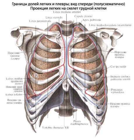
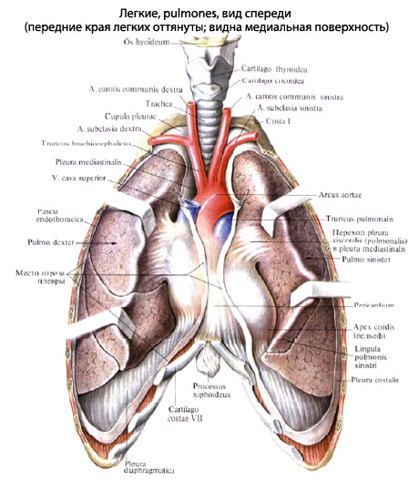
Lungs, pulmones(From the Greek - pneumon, stars fired legends - pneumonia), rots in the chest empty, cavitas thoracis, on the sides of the heart and the great ships, in the pleural bears, in the middle of the last median chest wall in front.
The right of the legend is a big obsyag, lower liv (by about 10%), at the same hour it’s very short and wide, in a persistent way, because the right dome with diaphragms is worth it to the left right of the part pechinki), and, in a different way, the heart grows more to the left, lower to the right, changing the width of the left legion by itself.
On the side of the snake, the lungs are lighter in the vascularized area of the cephalalus, as the vascularized gas exchange, at that time, the caudal, ale non-vascular saccular area is visible for ventilation. Two evolutionary paths to the development of the lung. In bronchoalveolar easy people function of ventilation and gas exchange to display thin, inflating alveolar tissue.
The essence of a bird's-eye light pole is in the fact that the ventilation works behind the help of non-vascularized children, which are even wider. It’s natural to be drawn into the bearskin’s mouth and then pumped up through the parabronchial area, like the day and the hard. A lot of gas exchangers are stored from blood-bearing capillaries, small enough to quietly, to take place in the lungs of patients, even if the large alveolar space is removed, the capillaries immediately adhere to even small capillaries up to 15 microns in diameter.
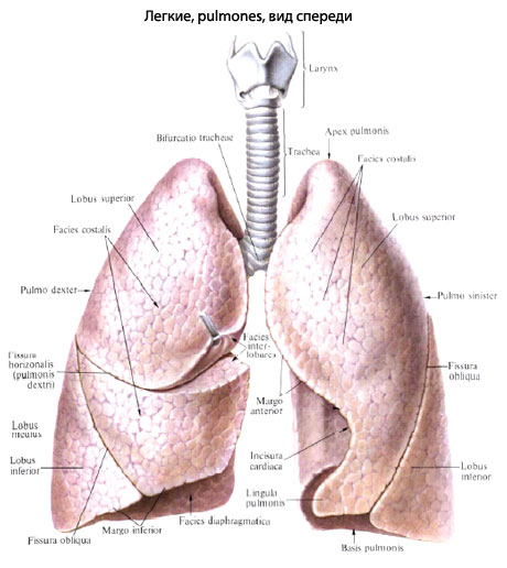
The skin is light, pulmo, we have an incorrectly cone-like shape, with the right side, basis pulmonis, we will straighten down, and with a rounded top, apex pulmonis, whistle 3-4 cm apart from the I rib, or 2-3 cm from the collarbone in front, behind and to reach to the level of the VII shyny ridge. At the top of the legend, there is a small bearded sulcus subclavius;
Tse descho asked to describe a bird's lung; napryklad, є kіlka of witches mіshkіv, and for the little birds є two sets of parabronks. Moreover, in the bird's lung є the gas exchange system is "perekhresy" with gas exchange;
As a result of the prominence of the projections of the partners and the enemies of the enemy. In the bronchoalveolar lung, inhale the gas is drawn in after the completion of the alveoli, and the thermal space is open for the mother to reach a large area of transverse transition for the diffusion of the natural gas for the expansion of the alveolar joints. There are one of the reasons why the alveoli in the legends of people are bigger, there are no capillaries in the legends of birds; the alveoli of the legends of people may have a diameter of about 3 mm.
An easy one has three surfaces. Lower, facies diaphragmatica The appearance of the swelling of the upper surface of the diaphragms is eliminated, to the extent that it is lying down. great Rebrova to the surface, facies costalis The swelling is due to the oppression of the ribs, as at the same time from the lying between them, the mid-ribs enter to the storehouse of the breast emptying.
In addition, the alveolar walls were to blame, they were even thinner, the stench could change its shape, and the gas penetrated into them along the way. As a result of the capillaries, thin alveolar walls become tight. Qia is a foldable structure є a direct inheritance of the reciprocating nature of ventilation.
The structure of the parabronchial lung is absolutely normal. Here, because of the gas є we have a single direct connection with the parabronks, the accumulation of gas diffusion for reaching the capillaries in the blood is less so, and there is no need in the great alveolar spaces of the lungs. Capillaries and capillaries of blood may develop approximately the same size. In such a rank, the structure of the parabronchial tissue is nagato mitsnish and igorstka.
Media surface, facies medialis, Rejected, repeated in a large part of the pericardium cut and extended to the anterior part, lay down to the middle, pars mediastinalis, and back, lay down to spinal stop, Pars vertebralis. The upper edges are visible: the gostry edge is given the name of the lower one, margo inferior; the edge, also a hospitality, is one of the fades medialis and costalis, - margo anterior.
Prove the rigidity of the parabronchial tissue and be seen from experiments; Such behavior strongly evolves from the one that is guarded in the legends of savts, if the parenchyma is forced to produce gas exchange as a result of the alveoli that are not ventilated or not. I know, the bird's light is carried in a tighter way, as the grip of the capillary moves. Tse zovsim is the best result, spasmed in the legends of the workers, if the occlusion is the same legendary artery make up razkogo fadinnya a support in the unsealed lung as a result of stretching and recruiting capillaries.
On the medial surface of the mountain and in the beginning of the death from the radix, the gates of the leg, hilus pulmonis, through the bronchi and the leg artery (as well as nerves) enter the lung, and two leg veins (and lymph nodes) pulmonis. At the roots lung bronchus to be drawn dorsally, the position of the legacy artery is not the same on the right and left sides.
The offense of chicks to the experiment is responsible for the nature of the parabronchial tissue. If I want the structure of the human lung, it’s good to go for its primary function of gas exchange, it’s easier and more inflated for other images, such as a vision. The difference is related to the fact that the thin alveolar tissue is responsible for both ventilation and gas exchange. Evolution has become more beautiful than rosetting in ptahs, de functions of ventilation and gas exchange from one to one. Apparently, apparently, the shortening design of a bird's lung in the meaning of the world is an academic interest for a pulmonologist, because there is no way of vicarious power in the lungs of patients.
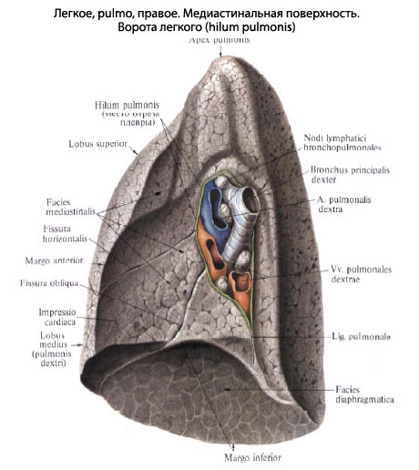
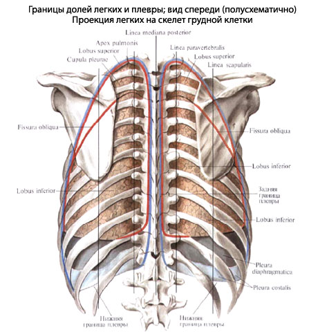
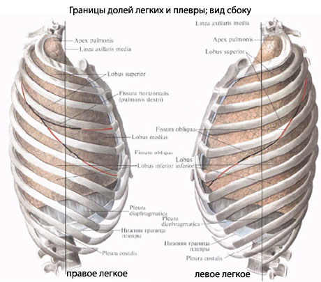
At the root of the right of the legend a. pulmonalis grows lower than the bronchus, on the left side of the bronchus it overflows and lies in the middle. Legendary veins on both sides of the root of the leg in the lower leg artery and bronchus. Behind, at the m_sci transition, one in one rib and medial surface a light, hostile edge does not endure, a part of the skin lung is rounded off to fit here in the buried chest emptyings on the sides of the ridge (sulci pulmonales). The skin is easy to help sore, fissurae interlobares, spread into parts, lobi. One beard, a scythe, fissura obliqua, scho can on both legends, to repair at a high rate (6-7 cm lower than the top) and then obliquely descend down to the diaphragmatic surface, more likely to go into the speech of the legend. Vona vidokremlyuє on the cutaneous lung, the upper part of the lower part. In the middle of the furrow, the right legend more than a friend, horizontal, furrow, fissura horizontalis, should pass on the IV ribs. There is a wedge-shaped dilenk from the upper part of the right leg, which becomes the middle part.
Kisen manages the process of energy, which will provide our clients with energy. If mi vidikhaєmo, mi viroblyaєmo dioxide in coal yak a by-product. Without a whole life of an important exchange, our children will quickly die and get overwhelmed, so they will get overwhelmed. Oskilki legeni blotted badly, stench є dinimi internal bodies, Yaki steadily poddayutsya in the influx of the most recent middle ground.
Our two lightweight tubes are stored in a folding grid of tubes, which are moved on the off-side of the heart, in the middle of the chest empty on the frame of elastic fibers. Once again, draw in through your mouth and nis, stay out of the way of the filter, and grab the particles of the saw on the hair. It’s better to warm up in front of him, like to go down the pipe, de vin breaks down to the bottom, between two dykhalny paths, called bronchi, which make it easy.

In such a rank, in the right legend there are three parts: lobi superior, medius et inferior. The legends have only two parts: the top, lobus superior, to which the top of the legend goes, and the bottom, lobus inferior, big volume, lower top. Until then, the entire diaphragmatic surface is visible and a large part of the back dull edge of the leg. On the front edge of the legends, in the lower part, the heart is viral, incisura cardiaca pulmonis sinistri. At the bottom, the tsya virizka is surrounded by a protruding anterior edge, called the tongue, lingula pulmonus sinistri. Lingula and lay down to her part of the easy view of the middle part of the right legend.
In the lungs, the lungs mucous bronchi split like the heads of a tree into tens of thousands of any smaller tubes, which are connected with the crumbly bears called the alveoli. The middle age is easy to place close to 600 mln. In one legend, fill the alveoli, cover the area, approximately equal to the area of the tennis court.
Alveoli - tse misce, de vidbuvatsya the most important exchange of gas. The little mice are soaked with a thin line of other blood-bearing vessels, or capillaries, as they come to the heart. Those, which are tied with legacy arteries, are the essence of deoxidized shelter, as it is necessary to innovate. Kissen pass through the neurally thin walls of the alveoli into the capillaries and then be carried back in the heart through the veins. At the same hour, carbon dioxide gas is seen from the blood through the same process of diffusion.
Budova legend. As a result of the submission of the lungs to parts of the skin from the two head bronchi, bronchus principalis, until the end of the legacy, repairs to parts of the bronchi, bronchi lobares. The right upper podovzhny bronchus, straight up to the center of the upper part, pass over the Legend artery and is called over-arterial; Some parts of the bronchi of the right legacy and all parts of the bronchi can pass through the arteries and are called podterialnymi. Parts of the bronchi, entering into the speech of the lung, see a number of other, third, bronchial tubes, which are called segmental, bronchi segmentales, as the stench ventilates the lungs of the lung - segments. Segmental bronchi, in their own way, are dichotomous (skin into two) into larger bronchi of the 4th and onset orders right up to endsevic and dichal bronchioles.
In fact, the robot doesn’t work in the main diaphragm, with a leaf of mouths between the breasts and black empty... It’s tricky to squeeze, since it’s dichotomous, wider lungs and tiny bittersweet. Mi just vidikhaєmo, weaken the diaphragm; easy to blow, like blowing cooler.
Light - a lot of fine organisms and irresistible for a whole series of ailments. Naybilsh extensions from them in the western regions є bronchitis and emphysema, which often vyklikayut kurinnya. Tubes in the middle of the lungs are chronically ignited, the excess slime is lost. The course can also lead to cancer of legends, head cancer, which is diagnosed in 4 million people in the river.
The skeleton of the bronchial vaschivaniya in a risky posture and in the middle of the lung of different minds mechanical injection on the side of the bronchus, the posture and in the middle of the organ: the posture of the lung, the skeleton of the bronchus folds from the cartilaginous infusions, and when it comes to the lungs between the cartilaginous infusions, there is a cartilaginous ligament, often in the structure of the lungs. In segmental bronchi and in the larger cartilage rosters, they do not grow into more form, but fall on the edge of the plate, the size of which changes in the world of bronchial caliber change; in the bronchial bronchial tubes of the cartilage. They get to know and sleep, and they have a little bit of a taste. The myazovy ball is stored in a circularly rostaty midi of the cartilages of the unscrewed myazovyh fibers. At the time of bronchial growth, special circular muscle bundles develop, which can sound or increase the entrance to the same bronchus.
Macro-microscopic Budov legends. The segments of the leg are folded from the secondary patches, lobuli pulmonis secundarii, which occupy the periphery of the segment with a ball up to 4 cm thick. Vona is adorned with semi-fabric partitions from the suspended secondary patches. interlobular available fabric on to take revenge on veins and lymph capillaries and sprites of crumbled patches in case of swelling of the leg. Even more often in nіy іvіdkladаєtsya vyhuvana vugіlny drank, as a result of the cordon, a piece of parcel will be clearly remembered. At the top of the dermal lobule, there is one bronchial (1 mm in diameter) bronchus (in the middle 8th order), to avenge the cartilage in its walls (lobular bronchus). The number of parochial bronchial tubes in the cutaneous lung reach is 800. The skin lobular bronchus grows in the middle lobules into 16-18 thinner (0.3-0.5 mm in diameter) bronchial bronchioles, bronchioli terminales, which do not replace cartilage and lobes. All bronchi, which are repaired from the head and terminated by the bronchial bronchioles, are stored in one bronchial tree, which serves to carry out the treatment during inhalation and vidihu; dyshal gas exchange between the witnesses and blood does not come into them. Kintsev bronchioles, dichotomously dry, give an ear of decal order of dichal bronchioles, bronchioli respiratorii; From the cutaneous dichal bronchial tubes to the radial enter the alveolar walk, ductuli alveolares, but end with the lame alveolar bears, sacculi alveolares. The skin wall of them is covered with a thick net of blood-bearing capillaries. Gas exchange is carried out through the wall of the alveoli. Dikhal bronchioles, alveolar walk and alveolar bears with alveoli store one alveolar tree, or dichal parenchyma of the leg. Pererakhovani structures, scho to resemble one of the bronchial bronchioles, set up a functional and anatomical unit, called acinus, acinus (grono).
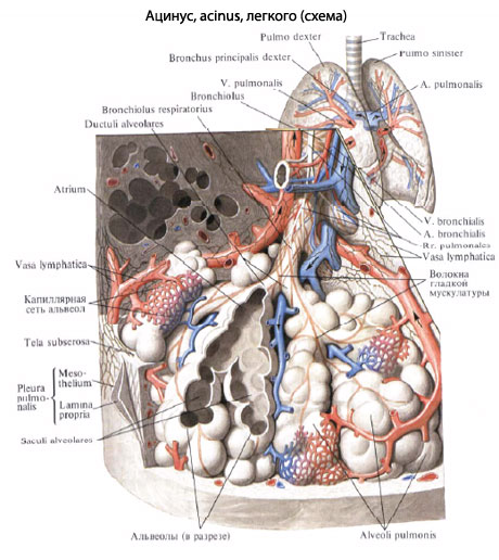
Alveolar walk and bears, to be carried to one dichal bronchiole in the remaining order, fold the primary lobule, lobulus pulmonis primarius. Їx is close to 16 in acinus. The number of acini in both legends reaches 30,000, and the alveoli 300-350 million. The area of the dysfunctional surface of the legends varies from 35 m2 with a vidih of up to 100 m2 with a large inhalation. From the suction of acinuses, there are small parts, from a small part - segments, from segments - lobes, and sometimes - more easily.
Lung function. The main function of the legends is gas exchange (souring blood and seeing it in carbon dioxide). Nadezhdenya in the legend of a rich sour drink and vivedennya to see, nourished in carbonic acid, it is called to be active dystopian rukhs chest wall and diaphragms and fast-feeling building of the lightest in the world from the range of mental health. At the same time, the speed and ventilation of the lower part of the river is great for the flow of the diaphragm and lower parts breast cancer At that hour, the ventilation and change of the upper part of the house will be the head of the rank for the help of the collapse of the upper part of the breast. The particularities give surgeons the ability to differentiate themselves to the retina of the phrenic nerve with visible parts of the leg. In addition to the extravagant dichotomy in the lung, the collateral dichotomy is developed, that is. It is possible to see through the pores in the walls of the legionic alveoli. In light older people, often in people with kidney disease, especially in the lower parts of the lungs, in order with partial structures є structural complexes, which are stored in the alveoli and alveolar passages, they are slightly spaced into the legacy parts of the acinus, and at the end of the day. The alveolar heights and allow the collateral dichotomy to function. So, as such atypical alveolar complexes tie around the broncholegenic segments, the colateral dichannel is not intertwined with each other, but wider wider.
The physiological role of the legend is not interchangeable with gas exchange. Їх of a folding anatomical attachment for displaying and developing functional manifestations: The activity of the bronchial wall during dyhanna, secretory-visual function, participation in the exchange of speech (water, lipid and salt with regulation of the chlorine balance), which is very important in the organisms. We will firmly establish ourselves, so that it is easy to volodynuyu with a forcedly developed system of cells, as it evokes phagocytic power.
Blood circulation in the legends. At the connection with the function of gas exchange of the leg, it is not only the arterial, but also the venous blood that can be removed. Staying through the arms of the legacy artery, the skin from which to enter the gates of the lung and because of the passage of the bronchial tubes. Finding the capillaries of the legacy artery is used to create a border of capillaries, which surrounds the alveoli (dichal capillaries).
Venous roof, flowing to the legacy capillaries through the legs of the legacy artery, enters into osmotic exchange (gas exchange) from the way in the alveoli: you will see your carbon dioxide in the alveoli and take away the acidity. The veins are stored in the capillaries, which provide shelter, they are sacked (arterial), and there are more venues in the storm. Remain angry at vv. pulmonales.
Arterial shelter is brought into the legends according to rr. bronchiales (from aorta, aa. intercostales posteriores і a. subclavia). Smell to liven up the wall of the bronchi and the legene tissue. With capillary hedgehogs, which pretend to be roughened in arteries, they are stored vv. bronchiales, scho run into chastkovo vv. azygos et hemiazygos, and sometimes in vv. pulmonales.
In such a rank, the systems of the legenevian and bronchial veins are anastomosed between themselves.
In the lungs, they develop superficial lymphatic vertebrae, embedded in the globular pleurisy, and gliboki, all the middle legenevia. The roots of lymphatic vessels are lymphatic capillaries, where they fix the holes on the respiratory and terminal bronchioles, in the interatomic and interlobular septa. The number of leggings trivializes in the gossip of lymphatic vessels near the hair of the leg artery, veins and bronchial tubes.
Introduce lymph judges and go to the root of the legendі lying here regional bronchogenic і distant tracheobronchial and nasal tracheal lymphatic universities, Nodi lymphatici bronchopulmonales et tracheobronchiales. So as to win the judges of the tracheobronchial nodes to go to the right venous cod, then the part of the lymph of the left leg is signified, going from the lower part, having been consumed in the right lymph duct. The nerves of the lungs are seen from the plexus pulmonalis, as if they were n. vagus et truncus sympathicus. Vyishovshi from the named gossip, the legene nerves expand in the parts, segments and parts of the lung along the bronchial and blood-bearing vessels, which can form the vertebral-bronchial bundles. In these bundles of nerves, gossip is established, in which microscopic internal nerves of the universities are perceived, preganglionic parasympathetic fibers are transferred to postganglionic fibers.
In the bronchi, there are three nerve gossip: in Advent, in the ball of the tongue, and in the epithelium. Pіdepіtelіalnom gossip reacha alveoli. In addition to the eferent sympathetic and parasympathetic energy, the afferent nerve is slightly unhealthy, as the bronchial tubes appear along the bloody nerve, and from the visceral pleural nerves - through the stores of the sympathetic passages of the thoracic nerve.
Segmental Budova Legen. The legends have є 6 tubular systems: bronchi, legeny arteries and veins, bronchial arteries and veins, lymphatic judges. There are a large number of cicatricial systems, one in parallel, one to one, fixing the supra-bronchial bundles, which form the basis of the internal topography of the lung. According to the subo-bronchial bundles, the cutaneous part of the lung is folded into the vicinity of the dylyanoks, which are called broncho-legene segments.
broncholegenic segment- the whole part of the lung, resembling the primary part of the bronchus and the supervascular part of the lung artery and the first part of the bronchus. The wind is seen from the middle segments in the bigger ones by the bends with the semi-tissue partitions, in which the segmental veins pass. Tsi veni with their pool half of the territory of the skin of the susceptible segments.
segments of the lung May the shape of irregular cones, for example, the top of the direction to the top of the lung, and from the top to the surface of the lung, sometimes between the segments, some of the growths in the pigmentation.
Broncholegenic segments are functional and morphological units of the lung, in the boundaries of which pathological processes can be localized, and most of which can be interchanged during the most agreeable operations to replace the resections of the lungs. I am very rich in classification of segments. Representatives of different specialties (surgeons, radiologists, anatomists) see a number of segments (from 4 to 12). According to the International Anatomical Nomenclature, in the right and in the legends there are 10 segments each.
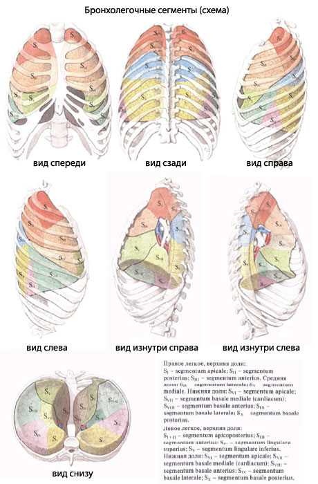
Name the segments of the given topography. Є such segments.
- Legend rights.
The upper part of the right legacy has three segments:- segmentum apicale (S1) borrowing the upper medial part of the upper part, entering into the upper opening of the breast cell and keeping the dome of the pleura; - segmentum posterius (S2) with its base of rectifying called і vіntsі, between them with II-IV ribs; the top of the yogo is turned up to the upper lobe bronchus; - segmentum anterius (S3) lying down before the anterior wall of the thoracic cell between the cartilages of the I and IV ribs; vin prilyagaє to the right anterior and upper empty vein.
The middle part has two segments:- segmentum laterale (S4) with its base of straightening forward і named, and the top - uphill і medially; - segmentum mediale (S5) stick to the front chest wall near the sternum, between IV-VI ribs; win prilyagє to the heart and diaphragms.
The lower part has 5 segments:- segmentum apicale (superius) (S6) borrows the wedge-like upper part of the lower part and grows in the paravertebral region; - segmentum basale mediale (cardiacum) (S7) by loaning the medical and diaphragmatic surface of the lower part. Vin prilyaga to the right anterior and lower empty vein; the segmentum basale anterius (S8) is located on the diaphragmatic surface of the lower part, and the side is large up to the chest wall in the groin area between the VI-VIII ribs; - segmentum basale laterale (S9) wedge in between the smaller segments of the lower part so that, from the beginning, it sticks out of the diaphragm, and the side of the side sticks to the wall of the chest wall in the groin area, between VII and IX ribs; - segmentum basale posterius (S10) of paravertebral sewing; lies behind all the other segments of the lower part, penetrating deeply into the posterior part of the costophrenic sinus of the pleura. One segment of the segment is seen as segmentum subapicale (subsuperius).
- Love the legend.
The upper part of the left legacy is 5 segments:- segmentum apicoposterius (S1 + 2) for the form and position of the seg. apicale і seg. posterius of the upper part of the right legacy. The front of the segment is slipped from the back slots of the III-V ribs. Medially, the segment is prone to the arch of the aorta and pidclavicular artery. You can also have 2 segments; - segmentum anterius (S3) є the greatest. I borrow a part of the costal surface of the upper part, between I-IV ribs, and also a part of the mediastinal surface, which can be attributed to truncus pulmonalis; - segmentum lingulare superius (S4) represents the dilyanka of the upper part of the III-V with the ribs in front and IV-VI - in the groin area; - segmentum lingulare inferius (S5) to grow lower than the upper one, but not to stick to the diaphragm. The offense of the lingual segment leads to the middle part of the right leg; stink of the heart, penetrating through the pericardium and chest wall into the costal-mediastinal sinus of the pleura.
The lower part of the left leg has 5 segments, As symmetrical to the segments of the lower part of the right leg, and that may be the same: - segmentum apicale (superius) (S6) borrowed paravertebral position; - segmentum basale mediate (cardiacum) (S7) in 83% of cases of ma bronchus, to be repaired with obstetric stubbur from bronchial tubes of the advanced segment - segmentum basale antkrius (S8) and medical surface of the leg; - segmentum basale laterale (S9) occupies the rib surface of the lower part in the groin area at the edges of the XII-X ribs; - segmentum basale posterius (S10) represents the great, spreading backwards from the other segments to the lower part of the legacy; it sticks to the VII-X ribs, diaphragm, lower aorta and stravohode, - segmentum subapicale (subsuperius) є not perceptible.
Innervation of the lungs and bronchial tubes. Afferent nobles from the visceral pleura breastfeeding cute stovbur, from parietal pleurisy - nn. intercostales i n. phrenicus, type of bronchus - n. vagus.
Eferent parasympathetic energy. Preganglionic fibers are repaired in the dorsal autonomic nucleus of the bloating nerve and go to the depots of the last and the third legacy to the plexus pulmonalis universities, as well as to the universities that have been expanded along the trachea, bronchial tubes and all the middle. Post-ganglionic fibers are directed from cych universities to the musculature and corpuscles of the bronchial tree.
function: sounding education of bronchial tubes and bronchial tubes and vision of mucus.
Eferent nice innervation. Preganglionic fibers go from the horns spinal cord upper thoracic segments (Th2-Th4) і pass through іdpovіdni rami communicantes albi і pretty stovbur to stellate і upper thoracic nodes. From the rest, post-ganglionic fibers are repaired, which pass in the storehouse of legendary gossip to the bronchial muscles and blood vessels.
function: expansion of bronchial education; sounding.
Here. Wonder in more detail about all the services of the clinic at the її.
As soon as you were before the boule viconani, Observe the results for consultation before the doctor. As soon as the viconan is not bully, everything is brutally necessary in our clientele, but in our colleagues in our clientele.
It is imperative to go further to the camp of your health as a whole. Є a lot of ailments, as a few things do not manifest themselves in our body, or in the bag to appear, but, unfortunately, it’s already nice. For the whole, it is simply necessary to develop the passes the rigging at the lykar, It’s not just a terrible ailment, but a healthy spirit in everybody’s body in general.
If you are supposed to be an organisation and a part of people, if you have a meal or a proposition - write to us, we will try to help you


