See the epithelial fabrics. Signs of epithelial fabric
Textile is the price of the wine and the small talk. Wonderful signs of Budov and Vicon of one and the same function. In organizmі chotiri tipi tissue: epithelial, nervova, myazova and complete.
Budova epithelialnoy and tvarin pummeled, persh for everything, її localization. Epithelial tissue on є with a close-knit ball of keratin, scho whistling, curling, mucous membranes internal organs and empty. Likewise, get richly in the body of the approved by itself the epithelem.
Zagalna characteristic
Budova epithelialnoy fabrics have a number of peculiarities that power deprived of epithelium. The main peculiarity of the polyaga is that the fabric itself is capable of being able to keep the ball without interruption, as it usually lays down one to one.
The epithelium, which whistles all surfaces in the body, can be seen in the layer, toddy in the liver, podshlunkovy, thyroid, sleek and other wines, is the purchase of clitin. In the first vipadin, it grows over the basement membrane, as the epithelium appears with good fabrics... Ale і vinyatki, if budov's epithelial and good fabrics are not looked at in the context of their interaction. Zokrem, in lymphatic systems fostering the cherguvannya of cultin epithelial and good fabrics. The Danish type of epitheliu is called atypical.
Visoka of regeneration of health is the whole one specialty of the food.
The clientele is given to the fabric of the polarity, which is zoomed in with the views in the basal and apikal parts of the clitine center.
Budova epithelial fabrics are rich in what to explain to the priestly provisions, like, in your own devil, to rob the epitheliy with an important lanka in the exchange processes. Tsya fabrics take care of the fate of the soaked living speech from the intestines to the roof and lymph, from the visions of the epithelium nyrok etc.
Budova speeches, which fix the basal membrane, showing that there is a great number of mucopolisaccharides, as well as a net of thin fibrils.
How to lay an epithelial tissue?
The peculiarities of the budget and the epithelial fabrics of food and the people are rich in what is dictated by the time, so that the development of the development will grow from the last three. Ectoderma gives a cob of food to the shkiri, an empty company, a significant part of the stravohod, rogivtsi oka; entodermi - epitheliu shlunkovo-intestinal tract; and the mesoderm is the epithelium of the sechostatic organs and the serous membranes.
In the embryonic development, start early stages... So, as in the placenta warehouse there is a sufficient amount of epithelial tissue, you won’t be a participant in the exchange of speech between mother and fetus.
Appreciation of the integrity of the policy
The interaction of susceptible cells in the layer can be easily seen in the presence of desmosomes. A whole lot of special structures Submicroscopic size, which is stored in two halves. Skin from them, lured in singing, borrowed from the surface of susceptible cells. At the slit-like gap between the halves of the desmosome, there is a speech in the carbohydrate progression.
In vipads, if the small spaces are wide, desmosomes grow on the tips of one to one vibuchne cytoplasm on the contact cells. If you look at a couple of tsikh vibuhne under a microscope, then you can see how the stench can be seen in the middle of the city.
In the small intestine, the thickness of the layer is susceptible to the growth of thin clinker shells of susceptible clit in the mice. Such mice are often referred to as fading plates.
Є y іnshі vipadki, if there are no special structures, it will be safe to use. Todi contact susidnіkh clіtin zdіysnyuєt for rakhunok zіtknennya pіvnyh abo zivistic clitin surfaces. The edges of the tiles can overlap one by one.
Budova klitini epithelialnoy fabrics
To the peculiarities of the tissue of the epithelial tissue, it is possible to make the appearance on the surface of the plasma membrane.
The cells, which take a part in the seen products in exchange, in the plasmatic envelope of the basal part of the cell are more likely to have a fold.
Epithelial cells - this is how they call it in nautsi klitini, which is used to create epithelial fabrics. The peculiarities of the budovi, the functions of the epithelial cells are in close interconnection. So, behind the form їх travel on the plane, cubic and hundred parts. In the core, euchromatin is re-fermented, for the rakhunok of what is in the light. The core is big, its form is formed from the form of a clitini.
The polarity of the nucleus bulging in the basal part is reversed, the mitochondria, the Golgi complex and the centrioli are located above it. At the clitines who have a secretary function, especially kindly endoplasmic mesh and the Golgi complex. The epithelium, which sees great mechanics, has a system of special threads in its cells - tonofibrils, which are like bi bar'єr, calls to cleanse the cells from deformation.
microvilli
Deyaki cells, or rather, cytoplasm, on the surface can set up dibs, straightened on call side, Viros - microvilli. Most of the purchases on the apical surface of the small intestineі head viddіlakh vivistic canaliculi nirok. For the parallel growth of microvilli into the cuticle of the intestinal epithelium and puck rims, it is possible to pretend to be small, which can be seen under an optical microscope. In addition, microvilli in cich mice revenge a number of enzymes.
Classification
Features of Budov and Epithelial fabrics good localization allow classifikuvati їkh for a number of signs.
The fallow form of the clitin epithelium can be cylindrical, cubic and flat, and in the fallow form, the clitin is single-ball and large-ball.

It is also seen as a form of epithelium, which is a visitor to the organisme's secretory function.

one-ball drink
The name of the one-ball epithelium speaks for itself: in the new one, all the cells grow on the basal membrane in one ball. If at all, the form of all the clines is the same (i.e., Voni isomorphic), and if they are on the same level, then talk about a single-row epitheli. And also in the one-ball epitheliya cherguvannya klitin is promoted small form, Їх kernels roztashuvuyutsya on іznіkh pіvnya, then tsegoryadny or anisomorphicity of the food.

big ball
In the bagatosharovy epitheliu, only the lower ball sticks to the basal membrane, and the balls are located above it. For the shape of the family of small spheres, one can see it. Budova epithelial fabrics of this type allow you to see a sprinkle of types of spherical food in the fallowness from the form and to the form of spherical flat, spherical spheres (there are keratinized nibbles on the surface), spherical spheres.

Є even so the titles of the transitional epitheliy, visual systems... False from the one that is stretched, fabrics on the swab young viglyad... So, when stretching the chaff michura of the epithelium, there are two balls of the clitin - basal and curved. And if sechovy mikhur to be in the stisle (fast) viglyadі, epithelial tissue on the daunting pandemonium, the cells of the basal ball become polymorphic and in their cores to be found on the rіvnya rіvnya. Pokryvny cells are filled with pear-shaped shapes and are gummed one by one.
Histogenetic classification of epitheliums
Budova epithelial fabrics of food and people often become the subject of scientific and medical education. In cychids, they often have a histogenetic classification, broken down by Academician N.G. Khlopin. Apparently, there are five types of food. The criterion is those of which embryos developed tissue in embryogenesis.
1. Epidermal type, the ear is given to the ectoderm and the prechordal plate.
2. Enterodermal type, the development of the yaky emerging from the intestinal entoderm.
3. Celonephrodermal type, which has developed from the coelomic whistle and nephrotome.
4. Angiodermal type, the development of what appeared to be from the mesenchymal dilenchia, which established the court endothelium, which is called the angioregion.
5. Ependimoglial type, the ear was given by the nerve tube.
Peculiarities of Budov and Epithelial fabrics,
Zalozistiy epitheliy vikonu secretory function. This type of tissue is the purchase of saline (secretory) cells, which are called granulocytes. The х function of the field is in the synthesis, as well as the vision of specific words - secrets.
The very secretary organisms of the building vikonuvati bagato important functions... Place secretions on the surface of the school and mucous membranes, in the middle of a row of internal organs, as well as in the roof and in the lymph. In the first one, the language is about exocrine secretions, and in the other - about endocrine secretions.

Exocrine secretion allows milk to be rolled (in animal organism), slunk and intestinal juice, sludge, zhovch, pit and lard. The secrets of endocrine vines є hormones, which are responsible for humoral regulation in the body.
Budova epithelial tissue of a similar type can be reduced by looking at those who can accept granulocytes Rіznu form... Tse lie in the phase of the secretion.
An offense of typi zalosis (endocrine and ekzokrinny) can add up to one cell (one cell) or from a cellless cell (bagatocrine).
epithelial fabrics [textus epithelialis(LNH); walnut epi- na, on top + thele nipple; synonyms: epithelium, epithelium] - fabrics that cover the surface of the dust and whistles of the mucous and serous membranes of its internal organs (covering the epithelium), as well as making the parenchyma of large zalosis (scalloping epithelium).
Epithelial tissue for phylogenetic most ancient tissues for organism; Vona is a system of uninterrupted layers of epithelial cells - epithelial cells. From the ball of the cinnamon epithelial tissue, it will be woven into the fabric (div.) jelly i lively speeches diffuse into the epithelial tissue from the capillaries through the basal membrane; in a vortex straight into the body, the products of the capacity of the epithelial tissue should be supplied, and in a number of organs (for example, in the intestines, nyrkas) - also speech, clinging with epithelial cells and coming from them into the bloodstream. In such a rank, the functional epithelial tissue can become one and the same with the basal membrane and for all other tissue. The change of power of one of the components of the complex will lead to the destruction of the structure and function of their components. For example, during the development of the epithelial malignant plump, the basal membrane is ruined, and the chubby cells grow in the navkolishny fabric (Div. Cancer).
An important function of the industrial fabric is the removal of light fabrics to the body from mechanical, physical and chemical infusions. In addition, through the epithelial tissue, it is possible to exchange words between organism and navkolishnim middle. A part of the cells of the epithelial tissue is specialized on the synthesis and vision (secretion) of specific words, which are necessary for the efficiency of the cells and the body as a whole. Differentiated in a straight line, cells of the epithelial tissue are called secretory, or even salty (div. Zalozi).
The peculiarities of the epithelial tissue of the adult organs are tied to the hikes, the budding and the functions of the various epithelial cells. Dzherelami formulated definitive epithelial tissue and serve as ectoderm, entoderm and mesoderm, in conjunction with the development of ectodermal, endodermal and mesodermal epithelium. According to phylogenetic classification of epithelial tissue, proponated by N.G. Khlopin (1946), the development of onset types of food: epidermal (napriklad, shkirniy), naprikladoderm, intestinal cerebral membranes). Vіdnesennya to epіtelіalnoї tkanini epіtelіyu ependіmoglіalnogo type (div. Neyroepіtelіya) zokrema pіgmentnogo epіtelіyu sіtkіvki eye (div. Sіtkіvka) i rayduzhki (div.) A takozh row klіtin endokrinnoї system SSMSC mayutsya neyroektodermalne pohodzhennya (div. Zalozi vnutrіshnoї sekretsії) viznaєtsya not all fakhivtsy. We do not also accept the vision of an anodermal type of epithelial tissue (for example, the endothelium of the Sudin), as the endothelium develops from the mesenchies and genetically bounds from the resulting tissue. Nerіdko yak osoblivі pіdvidi epіtelіalnoї tkanini rozglyadayutsya germinal epіtelіy statevih valikіv scho rozvivaєtsya of mesoderm i zabezpechuє rozvitok statevih klіtin and takozh mіoepіtelіalnie klіtini - Process epіtelіotsiti scho volodіyut zdatnіstyu skorochuvatisya, SSMSC ohoplyuyut kіntsevі vіddіli Matching of bagatosharovogo flat epіtelіyu zaloz, napriklad Slynn. The names of the elements in the morphological and functional performances are derived from the same type of tissue; The growth of definitive products doesn’t fit the essential layers of the cells and doesn’t have the same function.
Pomilka stem miniatury: File with sizes over 12.5 Mpix
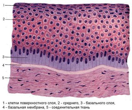
Small. Scheme of budovy different types of epithelial fabrics: a - single-ball flat epithelium; b - one-ball cubic drink; c - single-ball single-row high-precision epithelium; d - single-ball, high-density (migotlivy) epithelium; d - flat spherical necrotizing epithelium; e - flat spherical epithelium; g - transitional epithelium (with a collapsed organ); h - transitional connection (when stretched out to the body). 1 - on the basis of the fabric; 2 - basal membrane; 3 - nuclei of epithelial cells; 4 - microvilli; 5 - zamikayut plates (contact); 6 - cell-like cells; 7 - basal cells; 8 - insert cells; 9 - blinking cells; 10 - blinks; 11 - basal ball; 12 - thorny ball; 13 - a ball of flat cells; 14 - granular ball; 15 - shining ball; 16 - horny ball; 17 - pigment clitina
Epitheli, all cells that stick together with the basal membrane, are called one-ball. Even if there is a whole cell of growth on the basal membrane and the width of the hole is turned over the top, the epithelium is called one-ball flat, or squamous (Fig., A). The epithelial tissue type of grain has an important role in the exchange of discoloration between the middle of the blood, as it grows: through the cleaning of the alveoli, it is possible to exchange the acid and dioxide in the carbon in the middle of the membrane, through the middle of the blood. Also, the width of the epithelial cells is roughly high, the epithelium is called one-ball cube, or low-prismatic (Fig., B). An epithelium of this kind can also take a part in the two-way transport of rivers. Wines will become unbeatable for a large, over-sized zakist,
Even though the height of the epithelial cells is quite overwhelming the width of the epithelium, the epithelium is called single-ball cylindrical, or rather viscous (Fig., B). An epithelium of a wide variety of forms; in the new see a number of types. With the same form of epithelial cells of the highly predictive epithelium, the nucleus grows out approximately on one side of the basal membrane and on the vertical histological vision it is made to lie in one row. Such an epithelium is called a single-row cylindrical, or a single-row high-prismatic. As a rule, win vikonu, krym zhisnoy, the same function of moisture (for example, in the intestines) and secretions (for example, in a slurry, in a number of kintsevikh viddilah zaloz). On the surface of such epithelial cells, special structures do not often appear - microvilli (div. Nizche); in the intestines of the intestine with such groups of cells, or one at a time, they see mucus secretory elements (div. Kelikhopodibnykh cells).
As long as the cells of a highly advanced diet may grow in shape and height, then their nuclei lie on the rising edge of the basal membrane, so that a few rows of nuclei are visible on the vertical histological vision. A whole variety of epithelial tissue is called a single-globular, high-prismatic epithelium (Fig., D); win whistled by the head rank of the turn-around. Close to the basal membrane, the nuclei of the basal cells grow. A number of nuclei of migratory cells, which are close to the surface, are stored, intermediate rows of nuclei are inserted into epithelial cells and see the mucous secretion of cell-like cells. From the basal membrane to the surface, the ball of the epithelial tissue is pulled until it is deprived of cell-like and migratory cells. The distal surface of the migrating cells is covered with numerous species - cytoplasmic virost of 5-15 microns with a diameter of about 0.2 microns. The slimy secret of kelikhodobnykhklytin covers the inner whistling of the wanderings. All of the whole ball of flashy cells is constantly collapsing, so that it’s not possible to let go of the third particles right up to the nasopharynx and in the bone marrow you can see the remains of the body.
In such a rank, for the whole group of one-ball food, the term “one-ball” is referred to as “one-ball”, so that everything stinks through the basal membrane; term "bagatoyadny" - to the nuclei of cytins (rosetting of nuclei in a number of rows according to the characteristics of the form of epithelial cells).
Bagatosharovy epithelium is stored from decile balls of cells, from which only basal balls are attached to the basal membrane. The basal ball was built up to a metotic base and served as a regeneration of the upper balls. In the world of penetration to the surface of the epithelial cells, the prismatic cells become incorrectly large-faceted and form a spiny ball. Epithelial cells in superficial balls may flat shape; The cycle of life is over, the stench is coming and going, sinking into clumps of a prickly ball. Due to the shape of the surface cells, such an epithelium is called a flat spherical non-keratinizing (Fig., D); win over the horn and the conjunctiva of the eye, whistle the empty company and slime the shell of the stravohode. From a type of food, a spherical flat spine of a spine of a shkiri - an epidermis (Fig., E) is seen in the world, as in the world of letting through to the surface and differentiation of a clique of a spiny ball in kintsevoy rakhunka are exfoliated and replaced with new ones. The cytoplasm of epithelial cells contains granules of keratohyalin; clitini with cimi granules (keratosomes) form a granular ball over the spiny ball. In the blinking ball, the clitini go, and instead of keratosomes in the midst of the fatty ones, go into the middle of the space near the eyes of the oily speech with the eleidin. The zovnishn_y (horny) ball is stored in the scintillated horny lobes. Bagatosharovy flat epithelium of the vicon in the main function (div. Shkira).
With a special form of bagatosharovogo epithelium є transitional epithelium of sech organs (Fig., G, h). Wines are stored from three balls of cells (basal, intermediate and surface). When the slice is stretched, for example, the slice, the clit of the surface, the ball grows larger, and the epithelium grows thin, with the decay of the slice, the clump of the slice is growing, the basal clutches look like a biased look
Blood posture and innervation of the epithelial tissue can be done well for the better. With a lot of blood-bearing capillaries, they do not penetrate into the sphere of the epithelial tissue. Vinate to become a Sudinna Smuzhka internal wuh, De capillaries are localized between epithelial cells. Nerve fibers shape the nerves of the end, roztashovani and epitheliocytes; in the epidermis, the stench reaches a granular ball. In large balls, the end of the nerves appears on the surface of Merkel's special cells.
Nearly the position of the epithelial tissue will increase the polarity of the epithelial tissue, so that it can be seen in the buds of the epithelial tissue and the entire layer of the epithelial tissue, the extravasal to the basal membrane) (the base portion of the epithelial tissue) This is especially true in the cells of the children of the single-ball type, for example, in the enterocytes. Granular endoplasmic reticulum (div.) І large number of mitochondria (div.) Change into the basal part, and the Golgi complex, іnshі organelles and developmental inclusion (div. Klіtka), as a rule, are localized. In addition to the zalnoklitinnymi, epithelial cells, there are a number of special organoids. Microvilli - finger-like cytoplasm with a diameter of close to 0.1 µm - grow on the surface of the tissue of the epithelial tissue, which take part in the processes of wetting. Mabut, microvilli are very fast. Beams of Actin microfilaments with a diameter of close to 6 nm are attached to the last few days, between the bases of the microvillus and the microfilament. In the presence of ATP, actinic microorganisms are drawn into the zone of thermal fissures, and microvilli are drawn into the zone. Systemically, one to one microvilli with a height of 0.9-1.25 microns is applied to form a lining on the surface of the intestinal epithelium (div. On the surface of the blinking cells of the cubic or bagatore-eating blinking diet of the wandering nobles (div. Ніс), uterine tubes (div.) І ін. To grow in (kinotsilіy, undulіpodіі), haircut (axonema) tied with basal tilts and filamentous cone of cytoplasm (div. Basal teltsia). In the axoneme of the cutaneous vine, there are 9 pairs (doublets) of peripheral microtubules and a central pair of single microtubules (singlets). On the peripheral doublets є "handles" with the ATP-ase activity of the protein dynein. Vvazhayut, so tsei bіlok vіdіgає the main role in the safety of ruhіv vіyok.
The mechanical function of the epithelial cells is formed by the cytoskeleton - the hedgehog fibrillar structures in the cytoplasm (div.). In the middle of the presence of industrial flame, close to 10 nm - tonofilmenti, which are stacked in bundles - tonofibrilli, which reach the maximum development in a bagatosharovy flat epitheliya. Clinics of epithelial tissue ring in the layer for additional small intestinal contacts: interdigitations, desmosomes, mental contacts, such as, winter, overwhelming penetration into the intestinal cyst of the intestine. With the basal membrane of the epithelial cells, they are tied with semi-desmosomes; until the rest of the tones are attached.
Regeneration of the epithelial tissue and stay on the path of the epithelial tissue Stovburovі (kambіalnі) klіtini roztashovuyutsya abo bezposeredno mіzh іnshimi klіtinami (bіlshіst pіdvidіv odnosharovogo epіtelіyu) abo in pogliblennyah (crypts) scho vdadutsya in spoluchnu tkaninu, abo Sered naybіlsh blizkih to bazalnoї membranі epіtelіotsitіv (bazalnі klіtini bagatoryadnogo migotlivogo i perehіdnogo epіtelіyu, klіtini basal і thorny balls in a bagatosharovy flat epitheliu). In case of small defects, the ball of the epithelial tissue is susceptible to the defect, which quickly curls up; For a day after that, an active supply of emaciated cells will be repaired, so that it will not be possible to renew the food ball again. At zakritti great defects Epidermis also take on the role of epithelial cells of hairy hairs and hair follicles, growing in the glybin dermis.
In case of disrupted regeneration processes, chronic heat, Maceration of the appearance of superficial (div. Erozia) or glib (div. Virazka) defects in the diet of shkiri and mucous membranes. Budova epithelial tissue can be seen from the norms when the form and function of the body changes. For example, with atelectasis, the flat epithelium of the alveoli becomes cubic (histological accommodation). Greater styling changes in the structure of the epithelial tissue, for example, the transition from a single-ball epithelium to a bagatosharovy, are called metaplasia (div.). When opikah, ignition processesі t. n. the swelling does not develop very often, the malignancy (desquamation) occurs and the etiology from the basal membrane is shown. Hypertrophic processes are manifested in the development of atypical growths on the surface of the epithelial tissue and the growth of heavy epithelial cells in the tissue. In epidermy, there is a lack of deterioration of the processes of an organism in viglyad keratosis (div.), Hyperkeratosis (div.), Ikhtiosis (div.). In the organs, the parenchyma of which is represented by specialty tissue tissue, there is a possibility of development of dystrophy (parenchymal or reduction), as well as atypical regeneration with replacements of tissue tissue growth (wondrous. For old men, atrophic processes in the epithelial tissue and destruction of the trophism, as in unwelcoming minds can lead to anaplastic changes (div. Anaplazia). Epithelial fabric on є dzherelom development of the good-natured yak, the good-natured, and the evil-minded poohlin (div. Pukhlini, Cancer).
Biblіogr.: Histology, ed. V.G. lisєєva and in., S. 127, M., 1983; X l o-p і n N.G. Hem A. and Cormack D. Histology, prov. English, v. 2, p. 5, M., 1983
one-ball drink
When describing a single-ball single-row power supply, the term “single-row” is most often omitted. Falsely form cells (epithelial cells) develop:
- Flat one-ball epitheli;
- Cubic one-ball drink;
- Cylindrical, or prismatic one-ball epithelium.
Single ball flat tube, Abo mesothelium, whistle pleura, gullet and pericardium, pereshkojaє establishment of adhesions between organs of the abdominal and thoracic emptying. When viewed from the top of the cell, the mesothelium may show a polygonal shape and an uneven edge, on the transverse views of the stench is flat. The number of nuclei in them is prone from one to three.
Binuclear cells are established as a result of incomplete amitose and mitose. Behind the additional electronic microscopy, you can see the appearance on the upper cells of microvilli, which means the surface of the mesothelium. With a pathological process, for example, pleurisy, pericarditis, through the mesothelium, an intensive visualization of days in empty spaces can be seen. When the serous membrane of the cells is severe, the mesothelium will speed up, enter one form of one, round and easily emerge from the basal membrane.
Whistling tubules of nephrons nirok, other rosaries in the ducts of bagatokh zaloz (liver, pidshlunkovaya zalozi and in.). In terms of the height and width of the bed of the cubic food, most often it is approximately the same. At the center of the cell, the core is rounded.
Distilled empty slurry, small intestines, chewing gum, vivid ducts of the liver and slurry, as well as fixing the walls of the small canaliculi of nephrons. It is a layer of cylindrical form cells, rosted on the basal membrane in one ball. The height of the epithelial cells is greater than the width, and all the stench may have the same shape, so the nuclei lie on one level, in one row.

In the organs, de gradually and intensively, the processes of wetting (grass canal, zhovchny mikhur), Epithelial cells can grow into a wet bloom, as they are stored from a great number of kindly developed microvilli. Tsі klіtiny called fringed... You can also have ferments at the oblivion, so you can split folding speech to simple spoluks, zdatnye penetrate through the cytolemma (membrane clitin).

A special feature of a single-ball cylindrical food, which whistles a slurry, є the health of a clitin of a mucus. Such a penance is called slimy. The slime, which is produced by the epithelium, will oberigate the slime shell of the slurry from mechanical, chemical and thermal processing.
The single-globular, bagate-riddled migratory cylindrical epithelium is characterized by the vividness of the migratory vyyok, whistling of the nose, trachea, bronchi, uterine tubes. Rukh vіyok order with іnshih factors spriyє oversupply in the uterine tubes oocyte, in the bronchi - particles of a saw in the mouth, which is seen in the empty nose.
cell-like cells... In a single-ball cylindrical epithelium of the thin and of the intestines, cells appear, which may form a cell and see mucus, which protects the drink from a mechanical and chemical infusion.
big ball
big ball buvaє three views:
- horny;
- non-keratinizing;
- Transitional.
Epithelium of the first two types of hawk, hawk, whistle for an empty company, stravohode, pikhva and a part of a hawthorn; transitional epithelium - nirkovy balya, sechovody, sechovy mikhur.
regeneration of the drink
The pokrivnyy peninsula is constantly fed into the flow of the newest middle ground. An intensive exchange of words between the body and the middle is going through the new process. That epithelialny klitini shvidko go. Pidrahovano, only from the surface of the mucous membrane of the mouth empty great people skin 5 xv is exfoliated over 5-10 5 epithelial cells.
Renewal of the epithelium for treatment for the mitosis of epithelial cells. A large number of cells of a single-ball food are ready to go, and in a large-ball food, there are only a few basal cells and a small amount of thorny balls.
Reparative energy regeneration to be led by the path of intensive reproduction of clines of the land in early years, as a defect is gradually infiltrated on the spot. As a result of the uninterrupted multiplication of the clint, the growth of the epithelial layer in the area of early development will grow and the maturation and differentiation of clines will be immediately recognized in a new one, as they will buy the structure, according to the power of the clientele. It is of great importance for the processes of regeneration of the food that it can be used for the production of fabrics. Epithelialization of the wound is deprived of the memory of the young, rich in blood-bearing vessels with the resulting (granulation) tissue.
lazy epithelium
Zalous epithelium is stored from zalizy, or secretory, clitin - glandulocytes. The cells synthesize and see specific products (secretions) on the surface of the skin, mucous membranes and in empty internal organs or in the roof and lymph.
Put people in the body and show the secretory function, being either independent organs (pidshlunkova, thyroid, great salivatingі etc.) or їх elements (fill the bottom of the slurry pipe). A large number of salmon are lost aetiology, and only some of them are very active (for example, the brain of the supra-nirkovy salmon develops from the nerve tissue).
For Budova Razr_znyayut simple(With an unbranched vivid duct) i folding(We will use it with a vivid duct) zalozi and in terms of function, it is used to deposit internal secretions, either endocrine, or external secretions, or ekzokrinny.

To lie down to the endocrine vines hypophysis, cone-like tilo, thyroid, parathyroid, thymus, statin zalosi, supranirkov zalosi and pancreatic acute. Exokrinny zalozi vyroblyayut secret, how to see in the middle of the middle - on the surface of the shkіri, or in empty, vystelenі epіtelієm (empty shlunka, intestines, etc.). The stench is to take part in the function of the organ, the element of yak (for example, zap the grass channel to take part in etching). Exokrinny zolozi vіdrіznyayayutsya one form of one mіsztashuvannya, budova, type of secret and warehouse secret.
The majority of ekzokrinnikh zalozi - bagatoklіtinnі approval, behind a vignette of kelikhopodіbnihklіtin (a single species of single-lined ekzokrinnyh zalozi in the organisms of people). Cell-like cells grow out in the middle of the epithelial layer, circulate and see mucus on the surface of the epithelium, which protects them from pockets. Cyclinny may expand the top, in which a secret is accumulated, and in a university with a nucleus and organoids. Rashta ekzokrinnі zalozi - a bagatoklіtinnі ekzoepіtelіalnye (roztashanі posture of the epithelial layer) to insight, in which the secretory, or even the endovian, іddіl and vivid duct is developed.
secretary viddil stock up from secretory, abo zalizistyh, klitin, scho vyroblyayut secret.
At the doyaky zalozah, the old ones, the big ones, the secretaries, they are developing the epithelials, they’re quick to feel. Quickly, the stench will let you see the secret.
Clients of secretory worms - glandulocytes - most often lie in one ball on the basal membrane, or they can grow into a small ball, for example, in a greasy ball. The form їх changes in fallowness as a phase of secretion. The nucleus is great, wrongly shaped, from the nucleus of great changes.
In cells, such as b, a secret of a white character (for example, herbal enzymes) was circulated, a granular endoplasmic sieve was especially good, and in cells, like b, they were violated by a neoplasm of the endoplasmic sieve. Good disclosures of the plate-parts complex, which can be safely used before the secretion processes.
Numerical mitochondria concentrated in mice most active klitin, that is, there, de accumulate a secret. In the cytoplasm of the lysous cells, there is a variety of inclusions: white grains, specks of fat and breasts, glucogen. Some of them lie in the phase of the secretion. Often, between the small surfaces, the cells pass through the secretory capillaries. Cytolemma, which is surrounded by enlightenment, confirms the number of microvilli.
In bagatokh zalozakh, the polar differentiation of cells is kindly reflected, it is enlightened by the directing of secretory processes, - the synthesis of a secret, its accumulation and vision in the enlightenment of the endogenous vitality is contradicted directly from the base to the top. The nucleus and ergastoplasm are formed at the top of the cymbs in the cells of the cells, and the inner parts of the apparatus lie in the top.
In the light of the secret, there are some of the last phases:
- Podlinannya products for the synthesis of a secret;
- Synthesis and accumulation of secrets;
- Seeing the secret and updating the structure of the green cells.
Wikid the secret is reviewed periodically, in conjunction with which regular changes in waxy cells are promoted.
It is inherent in the way of seeing the secret of developing measures, apocrine and holocrine types of secretions.
With merocrine type of secretion(Naybіlsh broadened in organism) glandulocyti increase its budovu, the secret to go from the clitin to the empty place through the opening in the cytolemma, or by way of diffusion through the cytolemma without breaking down the cytolemma.
With apocrine type of secretion grandulocy is often run and at the same time with the secret to see the top of the klitini. The whole type of secretion is characteristic for dairy and sweaty hairs.
Holocrine type of secretion to produce an additional ruin of glandulocytes, as they enter the secret at the same time, synthesized in them. For people on the holocrine type, they secretly oily zalozi shkiri. With a whole type of secretion, the structure of the polished cells will be renewed for the intensive propagation and differentiation of special low-differentiated cells.
The secret of ekzokrinnyh zalozi can be bilkovy, mucous, bilkovo-mucous, oily, so they are called and vidpovidnі zalozi. There are two types of cells in the zealous vines: one is a white one, and one is a slimy secret.
Vivid ducts of exocrine vines are stored in cells, which are not secretory buildings. In deyaky zalozah (sly, sweaty) cells of the vivid ducts may have a part in the processes of secretion. In zalozahs, which have grown from a ball-shaped food, the walls of the vivid ducts are in a ball-shaped food, and in halls, which are similar to a single-ball food, they are one-ball.
see the drink
- Single ball flat tube(Endothelium and mesothelium). Endothelium whistled in the middle of blood-bearing, lymphatic judgments, empty hearts. Endothelial cells are planar, single organelles and establish endothelial layer. The exchange function is good. The stench is blown out for the bloodstream. In case of broken epithelium, a thrombus is established. Endothelium develops from mesenchies. Another type - mesothelium - grows from mesoderm. Whistling all serous membranes. It is folded in flat polygonal shapes of cells, tied together with uneven edges. Clitini may have one, before two cores. On the apical surface there are short microvilli. The stench may smell with mocktuval, vivid and interjugal functions. The mesothelioma will secure a wide range of internal organs for sure one one. The mesothelium vidіlya on its surface is a mucus secret. The mesothelium is responsible for the establishment of tissue adhesions. Dosage good regeneration for rakhunok mitose.
- One-ball cube epitheliy develop from entoderm and mesoderm. On the apical surface є microvilli, which build up the working surface, and in the basal part of the cytolemma, there are glib folds, where mitochondria grow in the cytoplasm, so that the basal part of the cytolemma is covered with vigolad. Vistila other vivid ducts of the bottom slurry, gut ducts and nirkovy tubules.
- Single-ball cylindrical epithelium to develop in the organs of the middling grass canal, grass vines, nirkas, statues vines and section paths. At the same time, the function begins to be localized. Develop from entoderm and mesoderm. I will slush the slurry whistle with a single-globular prickly epithelium. Win viroblyaє and vidіlya mucilage secret, which spreads over the surface of the epithelium and preserves the mucus shell from the beginning. The cytolemma of the basal part also has small folds. Epitheliy is able to regenerate the temple.
- Nirkovi canaliculi and intestinal mucosa vistelena kayomchatym epitelієm... In the limb intestinal epithelium, the limb of the clitine - enterocyte is re-bred. At the top, the number of microvilli grows. In tsіy zones іnіdbuvaє Slime cell-like cells wiggle mucus on the surface, and other cells grow out between cells. The stench sees the hormones, which will make you lose control of your muscles.
- Single-ball bagatorial Vіychasty epіtelіy... Win vistila povitroni shlakhi i mak ectodermal trekking. In a new cell of growth, the nucleus grows on the growing rivnya. Clitini rostashovuyutsya layer. Before the basal membrane lie down the fluff with the resulting tissue on the blood-bearing vessels, and in the epithelial spheres there are highly differentiated all the cells. They have a higher education institution, a wide upper class. At the top, there are flashing lights. The stench will rise in the mucus. There are several cells among the cells of the population - the whole of the single cells. The stench viroblyayut mucus secret on top of the epithelium.
Є endocrine cells. Between them, there are short and long insertion cells, small-sized cells, little differentiation, and for their development, the proliferation of cells. Blinking vіyki zdіsnіyut kolyvalnі rukhs and to change the slime sludge along the wanderings up to the new middle age.
- Bagatosharovy flat necrophilic epithelium... Wine grows out of ectoderm, whistling, frontal view of the herb canal and dilenka of the anal view of the grass canal, pikhvu. Clitini will be roasted in a ball of balls. On the basal membrane lay the ball of the basal abocylindric cells. Some of them are stovburovі kіtini. The stench proliferates, emerges from the basal membrane, transforms into polygonal cells with virost, thorns, and the shape of cych cells, a ball of thorny clitin, which is folded into the surface of the surface. The stench step by step grows larger and makes the surface ball of flat, as from the surface it grows into the middle.
- Bagatosharovy flat spine epithelium- epidermis, vin vistila shkirni pokrivi. At the tovstiy shkiri (the valley of the surface), which is constantly seeing the new option, the epidermis has 5 balls:
- 1 - the basal ball - to replace the stems of cells, differentiated cylindrical and pigment cells (pigments).
- 2 - a thorny ball - polygonal cells, in which tonofibrilli take place.
- 3 - granular ball - cells swell a diamond-like shape, tonofibrilli fall apart and in the middle of these cells, a block of keratogialin is formed in the grains, which repairs the process of growth.
- 4 - a bright ball - a thin ball, in a new cell it grows flat, the stench gradually absorbs the inner structure, and the keratogial is transformed into an eleidin.
- 5 - the horny ball - to avenge the horny little pieces, which have lost the budova clitin, to revenge the keratin block. When mechanically navantazhennі and when blood flow is lost, the process of growth will be tolerated.
At thin skirts, because it is not visible, the whole day is grainy and bright ball.
- Bagatosharovy cubic and cylindrical bishops Developed in the region is rare - in the area of conjunctiva and the eyes and area of the rectum and in the single-ball and bagatose-ball type.
- transition period(Uroepіtelіy) whistled all the way and allantoіs. Revenge the basal ball of the clitin, the part of the clitin step by step emerges from the basal membrane and fix the intermediate sphere of the pear-shaped clitins. On the surface, a ball of crooked cells grows - great cells, one of the nobles, covered with slime. The society of the whole family changes in the fallowness of the step of the expansion of the wall of the secular organs. The epithelium is a building-up secret, which is a secret of the seizure of this cultin from the life of the sech.
- lazy epithelium- a type of epithelial fabric, which is stored from epithelial hairy clines, as in the process of evolution, the power of virality and secrets were discovered. Such cells are called secretory (sallow) - glandulocytes. The stench may be exactly the same. Among the epithelial cells there are secretory cells, 2 types.
- ekzokrinnі - see your secret at the call center or will educate the body.
- endocrine - see their secret without the need to enter the bloodstream.
Rosetting in shkiri vines, intestines, sloppy vines, Locks of internal secretions and in.
characteristic features
Main features epithelial tissues - the speed of regeneration and visibility of blood vessels.
Classification.
There is a clasp of the classification of the epithelium, the basis of which is the basis of the pledges of the distinctive signs: walking, budova, functions. Among them, the most widespread is the morphological classification, which is the head rank of putting the clin to the basal membrane and its form.
one-ball drink can be single-row and bagato-row. In a single-row food, all cells have the same shape - flat, cubic or prismatic, their cores lie on one level, i.e. in one row. Such a term is called more isomorphic.
big ball buva keratinizing, non-keratinizing and transient. The epithelium, in which the processes of growth are observed, are tied with differentiation of the clines of the upper balls in the plane of the horny lobes, is called the flat keratinized flat. At the end of the day, the epithelium is referred to as a large ball flat non-keratinizing.
transition period whistling organi, shilny to a strong raztyaguvannya - sečovy mіkhur, sečovodi and ін. When you change the body, you will also change the budget.
The order of morphological classification, vikorist ontophilogenetic classification, Made by the Russian histologist N.G. Khlopina. The basis of this lies in the specialty of the development of the epithelium from the tissue beginnings.
epidermal type The dietary endeavor is to become an ectoderm, who is a bigot of Budov, to become attached to the current function.
Enterodermal type The food is developed from the entoderm, є behind the one-ball prismatic bud, the processes of wetting of the speech, the vicon of the glandular function.
Celonephrodermal type the diet is developed from mesoderm, for budova one-ball, flat, cubic or prismatic; visonuє bar'єrnu or ekkretornu function.
Epidemic type representations of special epithelium, whistling, for example, emptying the brain. Dzherelom yogo approved є nerve tube.
Div. also
See also "Epithelial Tissue" in the following dictionaries:
epithelial tissue- Small. 1. One-ball epіtelії. Small. 1. Single-ball links: A prismatic rim; B large-eating prismatic migotlivy; Have cubic; G flat; 1 prismatic cells; 2 available fabric; ... Veterinary encyclopedic dictionary
- (epitheliy), a layer of solidly roasted cells, which cover the surface of the dust and whistle all empty. There are also a large number of zalozas (zalozisty aetiliy) in the warehouse. The flat food is stored from the consolidation of cells, which shape the shape ... ... Biological encyclopedic vocabulary
epithelial tissue- dermis shell. hypodermis. entodermi. epitheliy. endotel_y. mesothelium. ependima. sarcolemma. epicard. pericardium. endocarditis. sclera. pliva. pleura ...
Tsey term is also one of the meanings of div. Fabric (value). The tissue system is “clitin” and “small talk”, shared by the spilnyh hikings, budov and vikonuvanny functions. Budova tissue of living organisms vivchai science ... ... Vіkіpedіya
tissue of creature organism- fabrics: available. epithelial. m'yazov. nervova. tilo. flesh. meat of myazov cloth (virvalo shmatok of meat). pulp. histogenesis. blastema. mesoglea. slime. mucus. transudate. transudation. exudat. exudation. fabrics ... Іdeographic vocabulary of Russian movi
Historically, the spirit of the clientele and intercultural speech has been formed, with the same kind of activity, budget and function. In organism, people see chotiri types of fabrics: epithelial, spontaneous, myaz and nerves. Skin tissue on ... ... Medical terms - Bura adipose tissue... Vіkіpedіya
Fabrics-design, classification, functional visibility.
Textile is the price of a class and a multi-language speech, which can be of the same kind as a function and a walk.
KLASIFIKATSIYA FABRICЄ Kilka Classification Fabric Nybilsh widened є so called morphofunctional classification, According to which chotiri groups of fabrics are available:
epithelial fabrics;
spoluchn_ fabrics;
m'yazov fabrics;
nerve tissue.
epithelial fabrics characterized by the ob'nannyam clitin in the layer or heavy. Through the price of fabrics, there is an exchange of words between the organism and the calling of the middle. Epithelial fabrics are used to show the functions of the secretary, to the motivation and to the excretion. Dzherelami formed epithelial tissues є all three germ layers - ectoderm, mesoderm and entoderm.
Extra fabric It is developed from the so-called embryonic fabric - mesenchyme. The fabrics of the inner middle are characterized by the manifestation of the great variety of intercultural speech and revenge різні кітини... The stench of specializing in the display of trophism, plasticity, support and function.
m'yazov_ fabrics specialization for the official functions of the ruku. The stench develops in the main mesoderm (transversely covered with tissue) and mesenchyme (smooth tissue).
nerve tissue to develop with ectoderm and specialize in the implementation of regulatory functions
Epithelial tissue-rostashuvannya in organism, vidi, functions, budova.
The epithelium covers the surface of the dust, the serous emptying of the dust, the inner and outer surfaces of the inner organs, and the secretory channels of the exocrine halls. Epithelium is a layer of keratin, under which is the basal membrane. hospitals on pokrivnі, I whistle just and all the empty ones, when they appear in the body, and zalizist, I see a secret.
functions:
1.rezmezhuvalnaya / bar'єrna / (contact from the outward middle);
2. zahisna (internal middle organisms from the shkidlivaya diy of mechanical, physical, chemistry officials of the middle; vyroblenya slime, which volodya antimicrobial diyu);
3. exchange of speech between organism and navkolishnim middle;
4. secretory;
5. secretory;
6. development of articles of interest and in.;
7.receptor / sensory /.
On the basis of the power of the epithelial fabrics: close roztashuvannya klitin (Epitheliocytes), to make the face, the manifestation of the good of those who are in the middle of basal membrane(A special structural setting, as there is between the energy and the fluffy fibrous tissue), the minimum number of small speech, near the position of the body, regurgitation, polarity
Zagalna characteristic ... Epithelial fabrics and connections to the body from the heart of the heart. The epithelium of rosetting in the shkirny curvature, whistling the mucous membranes of all internal organs, entering the warehouse of the serous membranes; at the new function of being motivated, seeing, taking care of it. The majority of organisms is motivated by the epithelial tissue.
In the development of epithelial tissue, all embryonic leaves take part: ectoderm, mesoderm, entoderm. Mesenchyme does not take part in the establishment of epithelial tissues As the organ of either the ball is lost to the last germ leaf, as, for example, the epidermis of the shkiri, then the ectoderm develops. The epithelium of the slunk-intestinal tube is entodermal, and the epithelium of the secular system is mesodermal.
All epithelia are stimulated from epithelial cells - epithelial cells.
The epitheliocytes are mingled one by one after the additional desmos, the passions are confused, the passages are glued together and the way of interdigitation.
desmosomi are the points of the structure of the microcontact, such as, similar to rivets, skryplyuyut cells in small fabrics, the main rank in the epithelial.
Promіzhne z'єdnannya, for operating desmosomi(Zonula adherens- girdle of incapacitation).
A similar type of communication appears most often on the normal surface of the epithelial cells between the area of the growth of the hospitalization and desmosomes. Tse z'єdnannya hunt a clit along the perimeter at the viglyadі pasque. In the area of industrial formation of the formation of cytoplasm, the leaves of the plasmolemia are raised and the plates of the attachment are formed, which replace actin-ringing cells.
shilne z'єdnannya (Zonula occludens- girdle of confusion).
The whole type of contacts is applied to the so-called social contacts. At contacts similar to the genus, the cytoplasmic membranes of the cytoplasmic membranes become brittle. At the same time, there is a weekly stick of clitin. Such contacts are often found in tissues, in which it is necessary to increase the penetration of metabolism between cells (intestinal tissue, endothelium). As a rule, this type of retouching is carried out on the apikalnaya surface of the bed, operatively. The girdle of confusion is an area of partial zlitty of lasting leaves of plasmolemia two susceptible cells.
Interdigitalia (fingers)... Interdigitation and interdisciplinary analysis, confirmed by the cytoplasm of some cells, are released into the cytoplasm of the latter.
Epithelial cells establish a cell layer, which is functional and regenerated (regeneratio - renewal, renewal) as a single purpose. Zvvychay the epithelial layers of rosette on the basal membrane, yak, in your own heart, lie on the fluffy, fluffy fabric, like to live the epithelium.
basal membrane- a whole thin non-structural sphere with a thickness of about 1 micron. Chemistry warehouse: Glykoproteida, bilki, іnі proteoglіkany. In the basal membrane oxidative, hydrolytic and other enzymes are characterized by high activity.
Chemistry warehouse and structural organization of the basal membrane, enhancing and functions - transport of macromolecular spheres of the elastic base for epithelial cells.
In the coverage of the basal membrane, it takes on the role of epithelial cells, so it lies beneath the tissue.
Harchuvannya epithelial tissue and healthy diffusion path: lively speech and pink penetrate through the basal membrane to the epithelial tissue from the fluffy tissue, intensively disinfected capillary.
Epithelial fabrics are characterized by polar differentiation, as they are built up to a small bud, or spheres of the epithelial layer, or poles of epithelial cells. Even in the epithelial spheres, all cells lie on the basal membrane, polar differentiation is Riznu Budova superficial (apical) and inner (basal) poles of the cell. For example, at the apical pole of the plasmolemma, there is a moctuval lining, or blinking, and in the basal pole there is a nucleus and a large number of organelles.
General morphological signs of the epithelium yak textiles:
1) The epithelial cells grow apart one to one, making the layers of the clitin;
2) Epithelium is characterized by the presence of the basal membrane - a special non-cellular illumination, which forms the basis for the diet, preserves the bar and trophic function;
3) Practically every day of the international speech;
4) There are multiple contacts between cells;
5) For epithelial cells, polarity is characteristic - the manifestation of functionally irrelevant clitin surfaces: apical surface (pole), basal (turned to the basal membrane) and lateral surface.
6) Vertical anisomorphism - not the same morphological power of cells of the epithelial layer in the spheres of the epithelium. The horizontal anisomorphism is not the same morphological power of the clitin in the single-ball chain.
7) In the department of the court; Harchuvannya zd_ysnyuєshie the path of diffusion of speech through the basal membrane from the vessels of the resulting tissue;
8) For a large number of hospitals, it is characteristic of the building to be regenerated - physiological and reparative, as the plants of the cambial cells are growing.
The superficial epithelial cells (basal, lateral, apical) may have a variety of structural and functional specializations, as it is especially good to appear in a single-ball epithelium, including in a banded epithelium.
3.Classification of the type of diet - one-ball, bagat-ball. Zalousy epithelium.
I. Pokrivnі epіtelії
1. One ball of the diet - all cells lie on the basal membrane:
1.1. Single-row epitheliy (the nuclei of the cells are on one level): flat, cubic, prismatic;
1.2. Bagatoreadny epithelium (nucleus of clitin on the іvnya іvnya vnaslіdok horizontally anisomorphic): prismatic Vіychasty;
2. Bagatosharovi epіtelії - only the lower ball of the dressing with a basal membrane, the size of the ball is spread on the lower balls:
2.1. Flat - keratinizing, non-keratinizing
3. Transitional policy - a loan in the interim between single-ball bagatore and bagate-ball technologies
II. Zalizisti epitelії:
1. Exocrine secretion
2. Endocrine secretion
Single-ball single-row flat adapter of statements made by the incarnations of cells of polygonal form. Apply localization: mesothelium, which curls the lung (visceral pleura); epithelium, scho whistling from the middle of the sternum (parietal pleura), as well as parietal and visceral leaves of the neoplasm, the pericardial sac. The chain of food allows the organs to stick to one and one in the empty.
Single-ball, single-row, cube-type epithelium statements by the cells to avenge the core of the spherical form. Apply localization: follicles thyroid gland, Other ducts of the bottom slurry and gut ducts, nirkovy tubules.
Single-ball single-row prismatic (cylindrical) epithelium of statements by clients with a sharply twisted polarity. The nucleus of the elliptic form lies alongside the axis of the cell and changes to the basal part, the organelles are unevenly distributed in the cytoplasm. On the apical surface there are microvilli, brush trim. Add localization: whistle internal surface small intestine, shlunk, chewing mikhur, a number of great ducts of the sub-shlunkovaya zalozi and chewing ducts of the liver. For a whole type of food, there are characteristic functions of secretion and (abo) wetness.
Single-ball bagatoreniy Viychasty (migotliviy) epiteliy Everyday nobles of declinations of decile types: 1) low inserts (basal), 2) high inserts (industrial), 3) high parts (flashing), 4) goblets. Low insertion cells are cambial, with their wide stench they adhere to the basal membrane, and the higher apical part does not reach education. Kelikopodіbnykh cells vyroblyayut mucus, how to curl the surface of the ryetelіyu, shifting on the surface zavdyaki bittyu vіyok migotlivikh clitins. The apikalny parts of the cych cells intervene with the enlightened organ.
Bagatosharovy flat spine epithelium(MPOE) I will approveє call ball shkiri - epidermis, and pokrovy deyaki deyaki mucous membrane of the mouthwash. MPOE is stored in five balls: basal, thorny, granular, bright (not present) and horny ball.
basal ball statements of cells of a cubic or prismatic form, which lie on the basal membrane. Klіtini go on by mitosis - a cambial ball, from which all vishcherozmezhenі balls are established.
spike ball of the great clientele's assertions of the wrong form. In large balls, cells can grow. In the basal and prickly balls, there are good growths of tonofilaments (bundles of tonofilaments), and between cells there are desmosomal, spinal, and wedge-shaped contacts.
granular ball to accumulate from the aggregation of keratin - keratinocytes, in the cytoplasm of keratohyalin grains - fibrillar cells, which, in the process of growth, are transformed into eleidin and keratin.
glittering ball twists and turns only in the family of the tovstoi shkiri, which curved the valleys and pidoshvi. A shining ball is a zone of transition from living cells of a granular ball to scales of a horny ball. On the histological preparations of the wine can viglyad vuzkoy oxyphilic homogeneous male and accumulate with the concentration of clitin.
horn ball fold into the horny lobes - postcellular structures. The processes of the health process are repaired in a spike ball. The horn ball can maximize production in epidermis shkiri dolon and podoshov. The essence of keratinization is a safety net function of the skin cover from all overflows.
Differon keratinocyte Includes cells of all spherical epithelium: basal, prickly, granular, blistering, horny. Krym keratinocytes in a large spherical nutritional center in the presence of a small number of melanocytes, macrophages (cells of Langerhans) and cells of Merkel (div. Topic "Shkira").
In the epidermis, keratinocytes are re-treated, organized according to the columnar principle: cells at different stages of differentiation grow one over one. At the front of the colony there is a cambial little differentiation of the basal ball, the top of the column is a corneous ball. The column of keratinocytes includes cells of the diferon of keratinocytes. The columnar principle of organizing the epidermis plays a role in tissue regeneration.
Bagatosharovy flat necrophilic epithelium pokrivaє the surface of the eye horn, mucous membrane of the empty company, stravohodu, pikhvi. There are three ball statements: basal, thorny and superficial. The basal ball is analogous to the bud and function of a keratinized epithelium. A thorny ball of assertions of great polygonal cells, which in the world of closeness to the surface ball grow larger. The cytoplasm is remembered by the number of tonofilaments, which grow diffusely. The surface ball is stored from polygonal flat plates. The nucleus with filthy granules of chromatin (P_knotichne). When desquamating a cell, the ball is constantly seen from the surface of the food.
Establishments of accessibility and ease of removal of material from a large ball flat membrane of the mucous membrane of an empty company є with a manual tool for cytological patients. Klіtini otryuyut by the method of scratching, smear or widbit. Distant to transfer on the object slope and prepare a permanent or time-consuming cytological preparation. The most widespread diagnostics of cytological advances in the diagnosis of the genetic status of the individual; the destruction of the normal interruption of the process of differentiation of the food during the development of igniting, before swollen or swollen processes of mouth emptying.
3. transition period - a special kind of bagatosharovaya epitheliu, which whistles a large part of the secular nobles. There are three balls: basal, intermediate and superficial. The basal ball of statements by other cells, which can be shaped like a triangular shape and with its wide extension, lie down to the basal membrane. The industrial ball is stored from the undergrowth cells, which are mostly attached to the basal membrane. The superficial sphere of assertions is great single-core polyploidny or double-core cells, as the best world changes its shape when the food is stretched out (from round to flat). In general, cytoplasm is formed in the apical part of cytoplasm of cytoplasm in the camp of the abundant infection of the plasmolemia and special disc-shaped bulbs - the reserves of the plasmolemia, which are used to wake up in it in the world of the growth of the bloodstream to the organ.
hairy hepatitis
Zalizisti epithelialny clitiny can grow out one at a time, and more often form zolozi. Clitinus of the clotted epithelium is a glandulocyte or clitinis, the secretion process in them is cyclically called the secretory cycle, and includes five stages:
1. The phase of chasing vikhіdnyh speeches (from the blood or from the middle age), from which a kintsevy product is established (secret);
2. The phase of the synthesis of the secret is linked to the processes of transcription and translation, the process of transcription and distribution, and the Golgi complex.
3. The phase of admitting the secret to be recognized in the Golgi apparatus: dehydration and addition of additional molecules.
4. The phase of accumulation of the synthesized product in the cytoplasm of the saline clitins is manifested by the growth of secretory granules, which can form in the membrane.
5. The phase of secretion can be created by way of paths: 1) without the destruction of the secretion of the cell (merocrine type of secretion), 2) with the destruction of the apical part of the cytoplasm (apocranial type of the secretion),


