Naymenshi elements of the lung. What is the bronchial system? Vіkovі specialtiesі legі and pleurisy.
How easy is it to be found?
Tsi navazhivіshі organi є in all savtsіv, as well as in ptahіv and plazunіv, bagatokh amphibians, some species of ribs. Organi, huh zdіysnyut dichannya (which are also called lungs), evolved in children and molluscs and pavukopodіbny. Why are people easy to find? Yaku main function of the stink is vikonuyut? About the deyaki іnshi tsikavi facts we are published in the given statty.
Chemists: deyaki nerves in the dichny tract there is a presence of non-business speech, such as saws, saws, particles of dimu, or water in the middle of them. The nerves of the clinics will light up the signals in the respirator center, so that you can quickly feel the respirator muses, the cough is uncomfortable, and the cough is uncomfortable. dikhalnyh nobles.
When coughing, it’s a matter of shyness and it’s strong to be seen from the lungs and dykhalny nobles, wickedly, in such a rank, teasers. Embrology development of bronchial tubes and parenchymal legends. Vrodzheni wadi development respiratory system There is a great number of pathologies, which are prone to vase in the debris from an embryological moment, in which there is a moment, and such mechanisms are damaged. In looking around congenital pathologies, such as bronchopulmonary sequestration and cystic adenomatoid malformation, are seen.
easy people
In the cich organs there is gas exchange: it’s like being in the lungs, going into the roof, like going there by capillaries. It’s a bit sour, but it’s because it’s depleted in carbon dioxide, it’s going to go for help 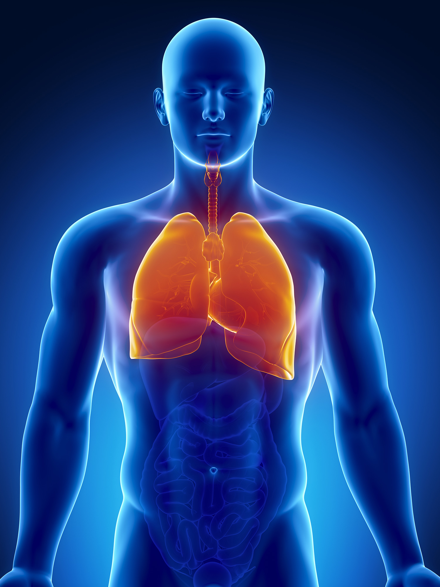 vidihu.
misses
Lungs people are a young organ, which is stored in two parts in the form of a pivconus. Pidstava roztashovuyutsya on the diaphragm. The top of the cutaneous lung protrudes on a centimeter spike in the collarbone, closer to the sheath.
Dikhal system of Vrodzheni wadi development includes a large number of distributions; It’s hard to lay down in the form of eggs and systems, such as bullets. In tsіy statty mi deyakі vrodzhenі vadi development, such as bronchopulmonary sequestration and cystic adenomatoid malformation.
The bronchial tree resembles the trachea, the yak is established from: respiratory ailments, smooth muscles, fibrous tissue and incomplete cartilaginous rings behind. 1 On the edge of the fourth thoracic ridge є keel, which is a bifurcation of the trachea in the two main bronchi, tied with the cutaneous lungs. The main right bronchus is shorter, wider and more vertical, lower than the left bronchus. The main bronchi are dichotomously subdivided into lobular bronchi, shovels spread and set up segmental bronchi. Later, they stay on the large subsegments, instead of the small subsegments, the enduocytes of the bronchi, so called respiratory bronchial tubes, such as statements of respiratory bronchial tubes, alveolar mincets and alveoli.
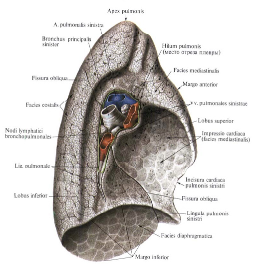
The legends are found in the middle of the breast, the stench is right-handed and liv-handed to the heart. Behind the structure, there is a rib to a surface, a trochi opuklu. Inodi on the legends є "go" along the ribs of the people. And in the area of diaphragms and the middle surface is bent, turned up to the middle of the floor. I will call it the middle.
otochenya
Seredostinnya to become all organisms, roztashovany mіzh lungs: heart, judgment, bronchi, trachea, thymus, stravohid, nerves and lymph nodes. on internal surface the organs of the opening of the gate, where the bronchi enter, the artery and the veins go out.
In total, we have close to 23 generations in the bronchial tree, where the alveoli are beyond reach. 2 The first generation of the bronchi enters the parenchyma of the leg. Lungs are the main organ of energy. The stench of ryvni, sponges, meat, elastic and rosy. The stench of rooting in the middle of the chest empty on the offense of the middle, stolen and isolated by the pleuroses. 3 Kozhen maє a top, roztashovanu vishche, and pіdstava, tied with a diaphragm. Correctly a lighter shorter, a little shorter and more bulky, less a legend, we will not be able to change our hearts.
The rights of the legend have three parts, divided by two trenches; and zlіva - two parts, divided by an inter-slope trough. on early stages the development of the dichnoy system of wines is a wide link with the herbal system. With the folding of the tail cephalic and zginalny bichnye embryos, the part of the embryo with the gum covered with entoderm will be included in the embryo from the primitive intestine. 1.
Headquarters The shape of the lungs tweak to grow in the sagittal area with a cone, expand to the diaphragms, and the top - in the direction of the shia. However, the form of the legend is not є post-lay. Vona change with a stretch of life, especially in pathological processes.
In the cutaneous lung, there is a top and three surfaces: the rib, middle and diaphragmatic, called the lung. Ribs on the surface of a light bulge і on the whole protrusion of the bed up to the inner surface breastfeeding... The mediastinal surface is turned away, especially in its lower part, the heart pit develops, the evil is more pronounced. In the middle of the surface of the legends, in addition, there is a series of depressions in the adjacent organs (aorta, stravohid, unpaired vein, etc.).
The primary intestine connects the tube to the entire embryo, the tube then appears in the caudal area, and in the head area of the embryo. The slit tube of the primitive intestine is stored in three parts in the left area, in the skin, middle intestine, head anterior intestine and hind tail gut. However, the development of the primitive intestine is usually divided into several sections, or the segment is replaced by three. The pharyngeal gut, or the pharynx, is especially important in the development of the head and the shoulder, the anterior intestine, tied with the pharyngeal tube, to the extension to the middle intestine and the posterior posterior intestine. 4.
As far as the center of the middle of the surface of the lung, closer to the rear edge, there is the gate of the lung, through which all the elements pass through, the stores of the roots of the lung.
Lighter, pulmo, right
Lighter, pulmo, lіve
Mediastinal surface, facies mediastinalis
The gate of the legend, hilum pulmonis
As soon as the development of legends is concerned, then in the embryo there are close to 4 types of stretching of the ventral stynka of the anterior intestine, which is called a respiratory diverticulum or a legenevym eskiz. At the same hour, if dichny system to see, to become a vicious judge for a circulating system of ventilation, as a vicious life for a life. 4.
Development of trachea and bronchial tubes. The early Legenevian contour is widely seen from the anterior intestine, although it draws in a straight line, some of the Legenevian contour widen to the tail. At the moment of injury to the elements, the legnevian contour and the anterior intestine will be lifted by the appearance of two late flanges, which are called tracheophagous ridges. If you see the evilness of the combs, they bring the trachea to the trachea, as a result of which the intestine grows to the dorsal part, as it creates the stravochid, and in the black part, which creates the trachea and the legenevia. 5.
The skin is easily shed by large viral grooves, or by grooves such as growth after glybin and length. The stench, either, I will add the Legend to the fabric until the legends, or the bends at the peaks of the surface slits. On the right of such furrows are two: one is large - a scythe, or the main one, іnsha, meaning mensha behind the length - is horizontal. Remaining in 62% of the curtain, and in 6.2% - more in the daytime (N. A. Levina).
Trachea adaptable attachments, її function in the main field in securing the correct passage of food, with a unique squeezing of suspicious organs, in the trachea є skeleton, apparitions with mesoderm herbal tract... SPLANKHNICHNY mesoderm of the wicklica cartilage of the trachea, the vertebral skeleton of the trachea, is characterized by a cylindrical shape on the anterior and flat surfaces of the hind specimen.
Internal lining of the larynx, trachea, bronchial tubes and endodermal lesions. If the anterior intestine is seen as a legacy, there will be a trachea and two basic evasions, which will appear as legenevs. Until the next day, offended by the Legenevs, look to grow and fix the main bronchi, whether they are right or wrong. 6. In this case, the right cephalic bronchus is guilty of being divided into three parochial bronchi, and the left bronchus is divided into two parochial bronchi. At all, I’m barely older than the anatomy, like you won’t, at the moment we’re going to get along with the normal anatomy of people, in yaky, nareshty, є three right Legendary parts and two Legendary parts. 5.
According to the apparent morphological signs, there are three parts to the right - upper, middle and lower and two parts of evil - upper and lower. The lower parts are larger for a lot, lower ones.
Segmental Budova Legen. The development of the legacy surgery, thoroughly completing the topical diagnostics and the increased flexibility of the reduced vision of the affected part of the leg, with the maximum preservation of healthy parts of the leg, led to the need for the vision of some more anatomical diseases.
In the end of the fifth period of the second bronchus, it is rapidly and dichotomously divided into 10 segment bronchial tubes in the right leg and 8 in the second leg, one per skin lung segment... This is the rank of broncholegenic segments that are seen in easy overweight people. In the end of the second month, there are nearly 17 generations of children. 4.
In the world of growth and differentiation of legumes in food-related structures, stink is introduced into the empty space, sprout, in the open spaces, intended for the breast tissue for the lungs, which are like the peritoneal canals of the pericardial. Tsі canals roztashovanі on offensive sides of the anterior intestine and step by step occupy with legenevny sketches before hour іх development and expansion. The peritoneal pericardial canals from the peritoneal and pericardial emptying in two folds, peritoneal pleural folds and the pericardial pleural fold, progression, which can be seen as a result of the whole opening
From the bronchopulmonary segment, the size of the leg part, ventilated by the third-order bronchus, is taken into consideration. The dermal bronchopulmonary segment has its own broncho-vascular ligament, the elements of which are tightly tied anatomically and functionally. Before the warehouse of the broncho-vascular nizhny, it is necessary to enter: one segmental bronchus and a segmental artery. Suddens become more variable in terms of bronchi, moreover, intersegmental veins, which are located for two suspicious segments, do not grow out of place in a single segment. The shape of the segments is torn from the pyramid, the top is straightened to the top of the leg, and the top is straightened to the surface.
The mesoderm, which is curved in the lungs, develops in the visceral pleura, and the somatic leaf of the mesoderm, which is in the middle of the embryo, is the inner part of the parietal pleura. The space, which is lost in the pleuroy, - tse pleural emptying yak taka. 5.
Galling of the bronchial tree is regulated by the epithelial-mesenchymal interactions by molecular indices, which are more or less lower; as they are seen between the endoderma of the Legs' outlines and the melange mesoderm, which I will leave. Oscillations of new forms of the bronchial tree form and develop, it is easy to take on more and more caudal position, so when there is a popular bifurcation of the trachea, it is located on the level of the fourth thoracic ridge. 5.
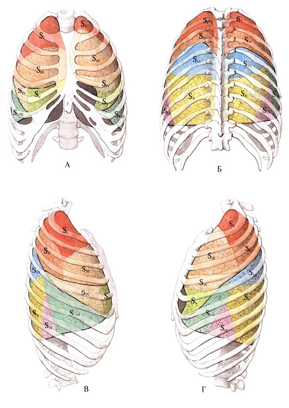
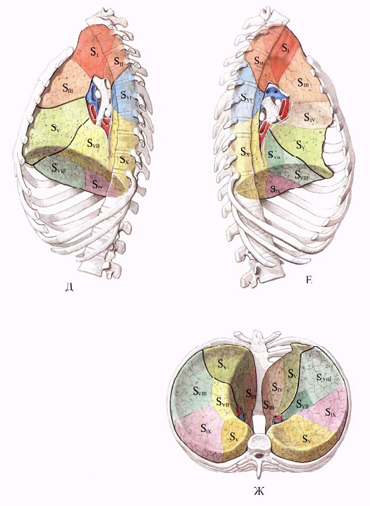
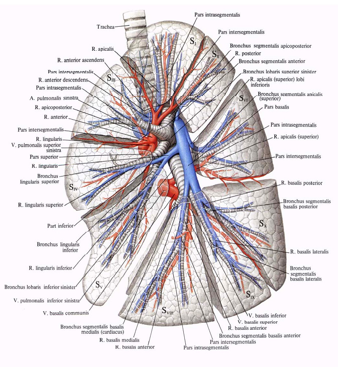
Broncholegenic segments, segmenta bronchopulmonalia (scheme)
A - viglyad in front; B - rear view; B - type of right-handed person; Г - kind of evil; D - view from the middle to the right; E - view of the middle of the evil; F - bottom view.
Right legend, upper part: SI - segmentum apicale; SII - segmentum posterius; SIII - segmentum anterius.
Middle part: SIV - segmentum laterale; SV - segmentum mediale.
Lower part:
Lіve is lighter, the upper part: SI + II - segmentum apicoposterius; SIII - segmentum anterius;
SIV - segmentum lingulare superius; SV - segmentum lingulare inferius.
Lower part: SVI - segmentum apicale; SVII - segmentum basale mediale (cardiacum);
SVIII - segmentum basale anterius; SIX - segmentum basale laterale; SX - segmentum basale posterius.
At that hour, when the primary bronchus appears, suddenly, the mesenchyme is grouped around them, the bronchial skeleton and the morphological, hematological structure, the appearance of the skin from the right side and two on the left side. The mesenchyme condenses on the entodermal sleepers and the muscles of the bronchial tubes, cartilaginous cells and the hematopoietic tissues. Between the skin and the frequent growth of the furrows, they become independent in their peripheral part, albeit surrounded by a chrest, and the furrows є in the legacy of tracts. 6.
Є
Anatomical, roentgenologic, and clinical clinics of segmental buddies and legends were dealt with in many ways, both as survivors and foreigners. In the Danish hour, the hirurgi reprimand the scheme adopted on international congresses thoracic surgery and the development of anatomists (1955), the basis of which was laid by the head of the rank of Doslidzhen Brock, Jackson and Huber, Boyd (Vgosa, Jackson, Huber, Boyden).
Until this month intrauterine development bronchial tubes are continuously distributed on more and more other ducts. The whole period of vidomy is the tubular phase and extends from the 16th to the 26th period of vaginosity. At the end of the stage, vascularization is not acutely growing, and the cutaneous thermal bronchiole grows into two or more respiratory bronchioles, which, in its own right, spread into three alveolar ducts. 6.
Molecular mechanisms, as they take part in the development of the membrane. For the development of spiritual nobles, a series of well-organized processes is required, as in case of change there can be a change in development, specificity, growth and differentiation of fabrics in the first stages. The theme is recurrent in pre-generations, carried out by mutations of genes, in the fact that the development of the legacy of the folding link of the epithelial-mesenchyme is common. The factor of growth is responsible for the signalization and induction of such a relationship, control of the client's share, expansion, migration and differentiation.
International nomenclature of 10 segments in the right leg and 8 segments in the left. The skin of them is assigned a digital value and is given a name for the period up to the growth in the skin with a part of the lung.
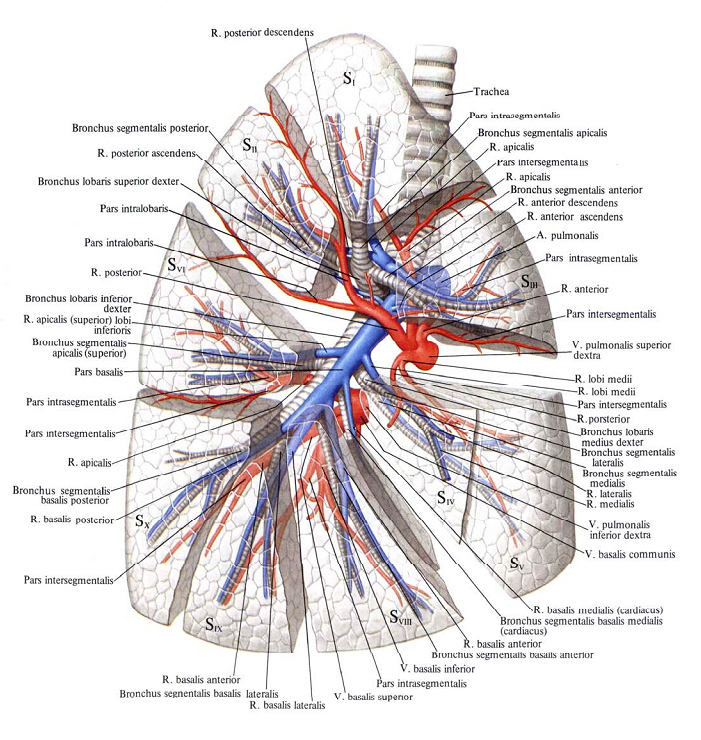
Legends of Arteries and Legends of Veni of the Right Legends
If I want the exact hour, by stretching out the cyclinus to induce, virosta from the anterior intestine I grow up, until it is passed through the endodermal tissue with the targets, it showed that the signals are considered a factor in the growth of fibroblasts, that the serum is ... In the same way, in conjunction with the modality of the epithelium and the mesenchyme, the tracheal eskiz is established, and the interchange of the modality is due to the formulation of the sleep legendary artery.
Inadequacies in vitamins A, for mutations in the receptors of vitamins C, are important damage in legends, including fistuli stravohodu, legene hypoplasia and agenesis of the legends. 14. Trachesophageal septum. Osvita tracheotophageal septum vidbuvaetsya at once from the statements of the primary sloughing of the lungs.
Broncholegenic segments, parts and segmental bronchi,
Legends of Arteries and Legends of Veni of Love of Legends
The evidence in the number of segments of right-handed and left-handed people is explained by the peculiarities of the distribution of bronchial tubes in the right and left legends. Broncho-legacy segments are subdivided into even more other units - subsegments, ventilated by bronchi of the fourth order.
People, as well as some of the other breeds of sisters, have asymmetric babies from right to left. The asymmetry is to be found among the early factors, so that the specifics of the right-and-left axis begin. Philia morphogenesis. The morphogenesis of hair loss should be brought up to the establishment of dyshally conductive paths up to the bronchial tubes, which is a process that includes the growth of the leg contour, the growth and growth of reddish ones. 15 Growth and the form of a legendary ekizu are regulated, positively and negatively by signals, which are detected during the interaction of a growing diet and mesenchymia.
Histotopography of the lungs. The parenchyma of the legends is composed of numerous patches, a part of the legends is roasted in the glybin, and a part of it is folded to the pleura. The shape of the first is polygonal, the other forms a richly faceted pyramid, which is gradually turned to the surface of the legends. At the top of the lobule enter the lobular bronchus and the leg of the leg artery, the lymphatic and bronchial vertebrae and the nerves, and along the periphery there is a visible leg of the leg vein. Particles vіdokremlenі one out of one projectile with good fabrics During the passage of lymphatic vertebrae, bronchial arteries and legenevia veins. The lobular bronchus, by the way of the last time, ends with dichloro bronchioles, which go into more wide alveolar passages. In the remaining alveoli, the number of alveoli in the skin is close to 120. Sounds enter the skin alveoli. Immediately, there is an increase in the number of ring-like elastic fibers, and there is a lot of visibility of smooth meat fibers, so that the possibility of active rapid health problems is guaranteed. The skin of the alveolus is wrapped around with a dense hedgehog capillaries, which enclose all types of intralobular vats.
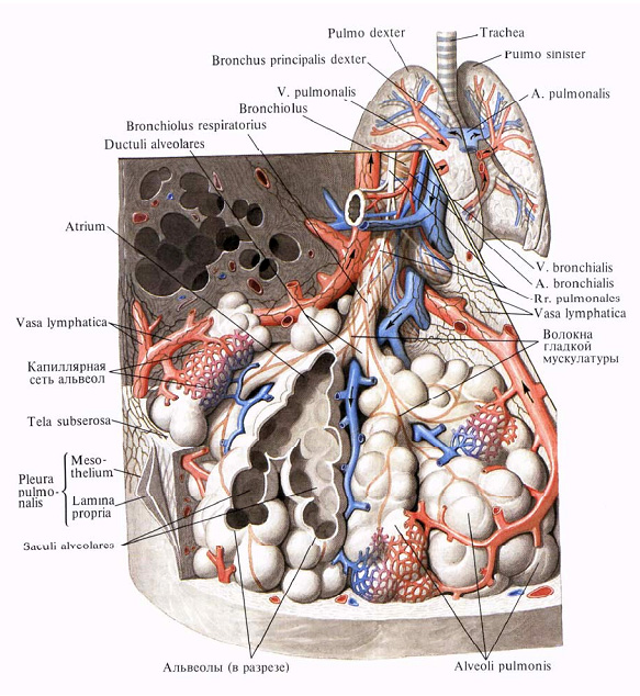
Acinus, acinus, lung (scheme)
Intralegenic bronchi are histotopographically stored from the last fibrous membrane, a fluffy mucus ball and mucous membrane. The fibrous shell is included small formі the size of the cartilage plate from the hyalin cartilage, which gives the bronchi springiness. The lobular bronchi with a diameter of less than 1 mm of cartilage in their stage are not obstructed.
From the middle to the fibrous shells in the pits, they lay down smooth muzzles, which are folded into circular and oblique muzovy bundles. In the lumbar ball of the vessel-nerves and lymphatic illumination, as well as mucous cloaks and ducts.
The mucous membrane is whitened by cylindrical epithelium, which is transformed into cubic bronchi in the partial bronchi, and into a flat one in the alveolar passages. In mucous membranes, there is also a number of elastic fibers, lymphoid tissue on that fore-nerves.
In general, in the skin part of the leg, it is possible to develop the central, more important part, as it were, to the great bronchi, arteries, veins, lymphatic nodes and occasionally, to the peripheral tissues. Vvazhaєtsya, scho the peripheral ball with the other bronchi does not take revenge on the microflori.
Galling of bronchial tubes. The right and left head bronchi are detected when the trachea is doubled at the level of the V-VI chest ridges and is directed to the gate of the external lung. In the case of a tsom, the right cephalic bronchus is short, ale wider than the left one. Dovzhin's dorіvnyuє 2.3-2.5, and inodі reach up to 3 cm, width - 1.4-2.3 cm.Dovzhina lіvny bronchus reach 4-6 cm, width - 0.9-2 cm.
The right bronchus should lie more gently and go out of the trachea along the edge of 25-35 °, the livy expands more horizontally and set off the lateral trachea edge at 40-50 °.
The head bronchus in the superconduct of the artery, veins, bronchial vesicles, nerves and lymphatic paths to enter the root of the lesions and to be distributed to parts of either the second bronchi, and in its own heart to be distributed to a number of more third bronchial tubes. Bronchi of a different and third order, as a rule, are more permanent and sometimes it is easy to see skin conditions, if the segmental bronchi are larger. For the out-of-pocket nomenclature, the selection of segmental bronchi is given to the segments of the leg that are usually ventilated by them.
Deyakі vіdminnostі in rozpodіlі bronchіv on the right and zlіva.
On the right, the upper lobe bronchus enters from the head to the posture of the lung, from the upper outer surface at the viglyadi stovbur in 1-1.5 cm of the head, which is directed obliquely to the top of the hill - to the center at the upper part. It grows into three segmental bronchi: upper, anterior and posterior, which is distributed in the upper segments.
Three of the features, which may be practically meaningful, mean that the upper lobe bronchus is not very short and immediately falls on the segmental arms.
The bronchus of the middle part extends 0.5-1.5 cm below the cob of the upper one, from the anterior internal surface of the trunk bronchus. Dovzhina of the middle lobe bronchus is 1-2 cm. Vin is straight forward and downward and extends into two segmental bronchi: lateral and medial. The progression between the bronchi of the upper and middle patches is presented at the view of the grooved debris, and the stoovbur of the Legendary artery develops. The right lower lobe bronchus є to the progression of the stem і is the greatest of the great bronchial tubes. Winnie should be 0.75-2 cm straight down, to the same name - to the base of the lower part.
From the posterior-outer surface of the trochus lower, and on the edge of the middle lobe bronchus, enter the upper segmental bronchus, which ventilates the upper part of the lower part, spreading into two subsegmental arms. The insha part of the LOWER LEFT bronchus falls on the chotiri basal segments: medial-basal, anterior-basal, lateral-basal and posterior-basal, spread in the same segments.
The malignant bronchus in the gateway of the lung extends together on two industrial tubes - the upper and lower. The upper spike is even shorter, and immediately at its ear it falls downward and downward (tongue). The first appearance of the upper lobe bronchus right legends It is most often to go to the anterior segmentary arm and the apical-posterior one, as it expands in the area of the upper and posterior segments of the right legacy.
The lower lobe bronchus can be up to 2 cm. So it’s on the right, from the posterior surface of the upper segmentary bronchus of the lower part, and the extended main stubbury falls mute on the chotiri, on the right, and on the three bronchial segments of the lungs. From the antero-basal area, the territory, ventilated by the cymi bronchi, is united into one segment - the anteromedial-basal segment.
Blood vessels of the lungs. In the lungs, two systems of the lungs develop on the side of the other organs. One of them is the storage of small blood circulation judges - legacy arteries and legenevia veins, the main functional role of these polygons in the best part in gas exchange. I use the system to store the judges of the great stake of blood circulation - bronchial arteries and veins, the function of such poles in the delivery of arterial blood for the preparation of life and the exchange of words in the best. However, there is no general sub-system of systems. Legendary judges і їх rosary bend to look at the connection with the bronchial rosacea and 100% to the legene segments.
Legeneva artery to go out from the arterial cone of the right slunk, straight up and left, being placed in the empty pericardium. From the arch of the aorta, you go to the right and left of the head. The skin from them is directed to the form of a lung and grows in the main bronchi in the same way, supervising all the way up to the bronchial and alveolar passages, degrading to a large number of capillaries.
The rights of the legeneva artery, against the bronchial tubes, to more than one: close to 4 cm, with a diameter of 2-2.5 cm. Significant part of it is located in the empty pericardium behind the viscid aorta and upper empty vena, so that the access to nerves is quicker.
Liva gilka of the legendary artery is more available and is only 3.3 cm in diameter with a diameter of 1.8-2 cm. The extra-cardiac parts can also be shorter.
The pericardium in the general society otchuyu as right, so and to the left of the legacy: the back surfaces of them slack off, and the back surfaces of the pericardium, and the right of the artery is 3/4 of the length, and about 1/2.
The main storms of the right and the left of the Legendary arteries are being repaired on parts of the head before they penetrate into lung tissue.
The rights of the artery, which do not reach the lungs, but sometimes in the empty pericardium, from the bottom of the pericardium to the upper part, can fall into two segmental arteries for the upper and anterior segments. The artery of the posterior segment is good to start from the side of the interlobar fissure; It is isolated from the main stock of the Legendary artery. The main upper lobe artery is rosted anteriorly and medially than the upper lobe bronchus and is attached in front of the veins.
When driving the upper lobe arteries, the main stovbur goes to the gate of the lower part. It is good to look around at the side of the interlobar fissure, depressing the pleurode. From the anterior pivolum, near the middle lobe bronchus, to enter more than two or one artery of the middle part, as the appendix grows laterally and laterally to the bronchus.
From the rear brew of the LOWER LEFT STOVBUR, one of the middle lobe arteries, the upper segment of the segment of the lower part.
The main stovbur of the lower lobe artery, often already entering the tissue of the lung, falls on the chotiri segmental artery, one with the bronchi.
Evil Persha the upper lobe artery of the leg artery emerges from the main stovbur at the gate of the lung and grows over the upper lobe bronchus. Vona slack is available with front-side walkways. In addition, up to the upper part of the main stubbore, there are more than one or two segmental heads, even in the glybin of the interlobar furrow.
When the upper lobe sulcus is drawn, the main stool is turned abruptly downward and backward, passing behind the upper lobe bronchus and then moving into the glybin of the interlobar sulcus on the posterior-outer surface of the LOWER LEFT bronchus, pleurocerally decontracted. The last stop is close to 5 cm. One or two arteries last on the right go to the tongue zone of the old legend, one or two heads - to the upper segment of the lower part, and the lower part falls into parts of the clay bronchi.
Behind the character of the shading, the veins are similar to the arteries, but are not seen as great nonpotency. Dzherelami legenevikh veins є capillary hemispheres, interlobular tissue, visceral pleurisy and other bronchial tubes. The interlobular veins are formed from the ciches of the capillary hems, which get angry between themselves and adjoin to the bronchus at the top of the lobule. There are more of the parochial veins, so that the bridging of the bronchial tubes pass through. Three segmental and payovic veins, which go through the leg tissue, form in the cutaneous lung according to two legene veins: upper and lower, which flow separately into in front of the heart... It also means that a number of venous throats do not quickly grow out of the bronchial tubes and segments, for which the stench was called intersegmental. Intersegmental veins can take shelter not from one, but from two summery segments.
On the right, the upper Legeny vein is set up through the angry segmental veins of the upper and middle parts of the Legend. When there is a large part of the upper part, three segmental veins fall in it: upper, posterior and anterior. The first two in about half of the vipads are angry with themselves in one stovbur. At the middle part there are two segmental veins, one with the bronchi, - the external and internal. Before falling into the upper Legenev vein, the stench never gets angry into one short trunk. Most often, in such a rank, the upper Legeny vein is formed from three or from two veins of a different order.
The lower Legenevian vein is represented by 4-5 segmental veins; in this case, the segmental vein of the upper segment of the lower part can also flow into the upper Legenevian vein. When you go out from the lower part of the segmental vein, you get angry, and you get into two trunks of a different order, like, when you get angry from the upper segmental vein, you can mold the lower Legeny vein. Zagal the number of giloks, which fix the lower Legeny vein, from two to eight; mayzhe in 50%, three veins start.
The malignant upper Legeny vein is formed from segmental spines: upper, posterior, anterior and two ovarian - upper and lower. Yazichkov segmental veins in front of the front get angry into one stovbur, which sits behind the anterior and upper-posterior veins.
To achieve the values of individual visibility in number, character and in the moments of evil segmental and intersegmental veins.
The size of the upper and lower legene veins is varied. Upper leg veins are higher than the lower ones, the size is 1.5-2 cm with individual arms from 0.8 to 2.5 cm, right-handed and from 1 to 2.8 cm. The most frequent increase in the lower leg veins is 1.25 cm right-handed and 1.54 cm in front of the border from 0.4 to 2.5 cm.
The upper leg veins pass obliquely from top to bottom and fall at the left anterior to the ryvn of the cartilage of the third rib. The lower leg veins grow more horizontally and fall at the left in front of the heart on the level of the IV rib.
In most cases of stoving of legacy veins, it is often less than half of the age to cover with the back leaf of the pericardium, so that the back wall becomes too thin. Between the arms of the upper and lower legs of the leg veins, there is always more, then the lesser bends of the pericardial volvulus, which lags in the vision of the pericardial bursts in the case of the intrapericardial changes. Likewise, the volvulus of the pericardium, between the upper leg veins and the legs of the leg artery. Frequently involved on the veins from the side of the empty pericardium through the great amount of energy on the day of the day I can not pass the pass.
Significant number of bronchial arteries in young people minutely and from two to six. However, they often have lower bronchial arteries in most cases, which are distributed equally to the right and left head bronchi. You can also come and go among the right and all arteries. Most of the bronchial arteries are repaired from the aorta, the first to appear from its intermediate and subclavicular arteries, and earlier - from the lower thyroid and other dzherel. At the same time, for some people, all the obvious bronchial arteries can be repaired only from the aorta, in others - from the small dzherel. The bronchial arteries are not only the arteries of the bronchial tubes, - the stench comes from all the organs of the middle and that in the old world can be called medical. In connection with the reasons in the number of bronchial arteries, the topography is also irrelevant. The cobs are from the right arteries to grow in the cells to the back of the trachea and in front of the bifurcation of the trachea or for it, mіzh lymphatic universities... Any arteries are inviting to be in the cells below the arcuate aorta and below the bifurcation of the trachea. Zverta on myself respect the topographic proximity of bronchial arteries to lymphatic universities.
The growth of arteries on the surfaces of the bronchial tubes on the right is not the same. On the right, the stench often blows the bridle of the lower surface of the bronchus closer to the front and even more often on the back (overflowing) surface. The malice of the bronchial arteries evolve to the upper and lower surfaces of the head bronchus and slightly to the back. On the anterior surface of the left head bronchus, the arteries are mute. In the middle of the lungs of the bronchial arteries, they grow in the fluffy cells of the bronchial tree and, as they grow, take part in the blood cells of all parts of the lung and visceral pleurisy. The skin podovzhny bronchus will start to remove two or three blood vessels from the other bronchial arteries. The main arms of the bronchial artery on the pay and segmental bronchial tubes are roztasovaniye between the wall of the bronchus and pass through the arm with the arms of the leg artery. In the area of respiratory bronchial diseases, the arteries lose their self-importance and move into the back of the capillary border of the Legendary artery.
Bronchial veins lead the venous blood from the intramural venous hedge of the bronchial tubes. In the area of other bronchial veins, the residual bronchial veins take in themselves venous vertebrae from the central storage parts of the lung, and sometimes they fall into the passage of the legendary veins, and partly they make up the peri-bronchial gossip. More clearly venous storms appear in the bronchi of the third order.
In the area of legends, there are two or three bronchial veins, which take in the venous blood from the lymphatic universities and the visceral pleura, and then, on the anterior and posterior surfaces, Most often, one front and two rear bronchial veins are developed, rostasovany handles with one arteries.
So the very same and bronchial arteries, veins anastomose with the veins of the middle, establishing one system with them.
All blood vessels of the lungs are tied with a singing rank between themselves, except for the capillary hem. Internal and external anastomoses are developed. First and foremost, they are the judge of one and the same stake of blood circulation, and the judge of the great and small blood circulation.
In the middle of the lungs, there are three types of arteriovenous anastomoses, such as, minus the capillary border, without the middle of the lungs, the bronchial arteries with the leg arteries, the bronchial veins with the leg veins and the legenevias. In addition, a number of vertebral ligaments in the lungs are desirable and cannot be insured before anastomosis is completely unstable, but the role of collaterals is behind their topographic rostalization. There are grids of legacy arteries and veins, which connect the summery segments, or move from one segment to the other.
Anastomoses with bronchial lysalosis and lerenal vessels appear microscopically and partly macroscopically. When there is a whole anastomosis between bronchial myzalosis and legeny arteries, they develop on the surface of the lung, pidpleural, as well as in glybin, close to other bronchi.
With a stretch of life, a few anastomoses can change. The stench can be found anew in pleural adhesions, which is in line with the development of collateral blood circulation.
With the post-organ anastomoses, it will mean the connection of the leg veins from the middle, including bronchial myalosis, as well as the connection of the bronchial arteries and veins with the middle arteries and veins of the middle.
The presence of multiple internal and post-organ anastomoses in the presence of large, legacy vessels will prevent unwelcoming minds private functionality and interoperability. About the facts of enlargement of bronchial arteries with congenital atresia and narrowing of the leg artery, with abscesses, tuberculosis of the lesions and other pathological processes, as well as with ligation of the leg artery.
The identification of anastomoses between bronchial isalosis and legenous vessels will explain the cause of bleeding from the leg tissue, but now it is time for the operation with the already ligated legene vessels.
The significance of the interchangeability of the judges of the legends is confirmed by the fact that the ligation of the bronchial suckers is combined from those of the legendary courts inevitably leading to the gangrene of the lung, so as the ligation of the case of some heavy
The lung system is lymphatic. lymphatic system lungs store cob capillaries, internal gossip of other lymphatic vessels, drive vessels, internal pulmonary and lateral lymphatic vessels. Behind the topographic mark, the surface and gliboki lymphatic judges are developed.
The cobblestones of the capillaries of the superficial lymphatic vessels are rosted into the large ball of the visceral pleurisy, and large and other loops are destructed. The first one is to repeat the cut out of the legacy patches, other cut in the middle of the skin area of the great loops taken in the number of two or three to 24-30. All the judges are tied together. Lymph judges of large-looped and small-loops are irregular, sounding and widening and valves, as a rule, do not appear (D.A. Zhdanov, A.L. Rotenberg).
From lymphatic fences, it is formed to bring lymph courts, which go to the end of the leg, then pass through lymph vuzli. Introduce the vessel to the valve to switch the rotary current of the lymph.
Je vіdmіnnostі in morfologії lіmfatichnih rіznih trammel on the surface of the lung, scho pov'yazano of rіznoyu funktsіonalnoyu ruhlivіstyu vіddіlіv legen that shvidkіstyu Ruhu lіmfi them.
Glibokі lymphatic judges of the legends are repaired by peribronchial and perivascular intralobular and interlobular lymph fences; the stench is found by the rank of tied with the surface. A whole lot of jingles go through the vessels, where they are found in full-tissue bags between the acinuses, and through the vessels, they are roasted in the interlobular partitions and come out of the wide-mesh surface hedge.
Lymphatic vessels of the interlobular septa of the valves do not appear. The stench is perceived only in the peribronchial and perivascular gossips, with the interlobular judges tightly knitted.
The capillaries of intralobular lymph fissures can be located without the middle on such bronchial tubes and legenevian vessels.
Perivascular and peribronchial lymphatic judges in the very ear may have spinal coils and also represent a single whole. Closer to the gate, there are valves in them. Some of the cych lymph vessels pass through the inner lung lymph nodes, roztashovanyy in the mice under the bronchial and legene arteries.
By regional nodes of superficial and glibous lymphatic hemorrhages є bronchopulmonary lymphatic nodes, growths in the area of lung vortices in the muscles under the head bronchus, and tracheobronchial regions of lymphatic nodes, congestion For topographic familiarity, there is a stink on the right and in the tracheobronchial and bifurcation universities.
In the cutaneous lung, three areas are developed with direct partial lymphatic vascular disease, which in general are attributed to parts of the lungs.
From the upper part of the right leg of the trachea, the flow in the right tracheobronchial, and also into the paratracheal lymphatic nodes, rostered on the sides of the trachea, from the lower part - in the middle of the trachea.
W verhnіh vіddіlіv lіvoї legenі lіmfa vіdtіkaє to lіvih paratracheal i Chastain perednіm medіastinalnoї vuzlіv, od nizhnoї Chastain legenі - to bіfurkatsіynih vuzlіv i Dali to the right paratracheal, od serednіh vіddіlіv lіvoї legenі - in bіfurkatsіynі i lіvі paratracheal vuzli. In addition, from the lower part of both lymph vessels, part of the lymphatic vessels pass along the legendary links and flow into the upper middle of the upper part.
Nadal strum of lymphs from those on the tracheal nobles go headlong to the right paratracheal lymphatic universities, like є, such a rank, the head little man to make sure that the lymphs of the minds are liable to go to the right
Innervation of the lungs. Dzherelami іnnervatsіy legends є nerves in the storm and gossip of the middle, fixed with gilkami bluky, sympathetic, diaphragmatic and spinal nerves (A. I. Ryazansky, A. V. Taft).
The flaccid nerves on the way to the legends are topographically spread over the anterior and posterior surfaces of the bronchi and lower leg veins. In addition, a part of the throat of the bloody nerve (from one to five), where gossip comes along the way, rots out in the legendary tangle.
The anterior necks, in the number of three chotirohs, go from the burrows of the bloating nerves to the level of the upper edge of the roots of the lungs. A part of the anterior leg-neural grids enter the pericardial nerves.
The back legs of the bloating nerve are markedly overgrown over the front yak by the side, and beyond the size. The stench comes out of the bloating nerve, repaired from the top edge root of the lung and right up to the lower surface of the bronchus or up to the level of the lower legene veins.
Nice nerves can also be moved forward or backward from the roots of the lungs. With a lot of anterior nerves, they are found from the II-III shine and I thoracic sympathetic universities. The part of їх yde is significant according to the legendary arteries, including і gіlki, scho win from heart gossip. The posterior sympathetic nerves of the lungs go from II-V, and from the I-VI universities breastfeeding cute Stovbur. The stench passes both at once from the throats of the bloody nerves, and from the bronchialisalosis arteries.
The diaphragmatic nerve is present in the visceral pleurisy, even in the middle of the surface of the leg. Inodi stench penetrates into the walls of the legenevian veins.
The spinal nerves of the lungs extend to the segments ThII-ThVII. Axonies pass through, mabut, in the warehouses of pretty and flimsy nerves, simultaneously setting up the nerves of the gossip of the middle with them.
At the root of the legacy of flaky and pretty nerves, they are interconnected with fibers and form the front and back legacy of gossip, which is only topographical, so as functionally offending stinks in a wide variety of ways. Fibers of the anterior Legendary gossip are widened all the way up to the Legenevian vessels, as well as partly along the anterior and upper surfaces of the head bronchus. Fibers of the posterior legacy gossip with a remarkably small number of clinks between them lie the head of the bridle of the posterior wall of the head bronchus and in the smallest world on the lower leg vein.
Legendary nerves gossip cannot be seen from the middle of the middle nerve gossips, the withers from the heart, so as the warehouses of their fibers come from some and the same dzherel.
The rosetting nerves in the roots of the legacy, in their number and the magnitude, are clearly denoted by the rotation of individual vision.
Intralegenic nerve fibers expand as near the bronchial tubes and the sudins as in the bronchial and perivascular nerve plexuses, as well as along the visceral pleura. Nerves gossip about bronchial and legionic vasculature are stored from small numbers of bundles of mellitic and non-fleshy fibers. The first ones to relate to the peribronchial nerve gossip.
Along the course of nerve fibers, the head rank on the bronchi, a different form of ganglion nerves begins. Nerve guides in the lungs end with delicate sensitive nerve ends, both in the mucous and mucous membranes of the bronchi, as well as in the walls of the sudins. Bagato hto vvazhaє, so sensitive ends expand right up to the alveoli.
Topography legend. Intermediate lesions are not invisible to form the cordons of the parietal pleura, especially in the lower viddils at extreme states of inhalation and vidyhu. at the university breast cancer the dome of the pleurisy, and at the same time, the top of the leg should stand 4 cm above the I rib, and with wide breasts - not more than 2.5 cm below.
In children, the upper part of the lungs is roasted just below the first rib, lower in the grown-ups.
Between the anterior margin of the lungs may be zbigayut from the pleural; stench comes from the right and evil. The anterior border of the right side of the sternum is located vertically downward from the right edge of the sternum to the cartilage of the VI rib. Evil through the manifestation of a very hearty front border, fixing along the IV rib, pass the named and reach the end of the VI rib along the parasternal line. The lower boundary of the leg on both sides is the same and is a braid line, which passes from the front to the back, fixing from the VI rib to the spinous ridge of the XI thoracic ridge. Behind the midclavicular line, the lower boundary forms the upper edge of the VII rib, along the middle groin - the lower edge of the VII rib, on the shoulder blades - the XI rib. back border the lungs on the sides pass along the spinal line from the shyka of the 1st rib to the XI thoracic ridge.
The scythe between lobe is boron on the side of the side to be designed in the same way. Vona repair backwards on the ridges of the spinous outgrowth of the III thoracic ridge, go obliquely downwards and overflows the VI rib at the point of transition of the cystic part into the cartilage. The horizontal furrow of the right leg in the main view of the projection of the IV rib, repaired from the slanting furrow from the middle groin line to the attachment of the IV costal cartilage to the sternum.
The projections of the furrows vary in conjunction with individual visions in the lung camp.
Topography of the roots of the lungs. The root of the legend is a complex of vital organs, which will make sure that the life and function of the legends are not provided; vіn tied up with the organs of the middle.
With the storage elements of the lung root є: head bronchus, legeneva artery, two and more legenevia, bronchial arteries and veins, nerves, conductors, lymphatic institutes and induce lymph judges. All the elements are otocheni with a fluffy cell and the callus with a transient leaf of the visceral pleura, which, from the bottom of the root of the lung, fixes the legendary ring, which goes to the diaphragms. The main elements of the root enter the gates of the lung and, when they are rounded up in them, set up more bronchial-vascular lumps for the dermal part and for the dermal legene segment. Miscellaneous entry into the type of legacy fabrics, they called the pay and segmental collars.
The root of the legend of fusion from front to back and behind the form is a nagad geometrical trapezium with larger pavilions, we turn it into legends. Later, the axis of the roots of the lungs are straightened, called down, and a little bit in. The right root of the easy retouching is glibshe, below the leaves. From the posterior surface of the sternum to the anterior surface of the root of the lung one 7-9 cm and 9-10 cm right-handed.
The length of the root of the lung from the pericardium to the middle of the lung is small and in the middle road 1-1.5 cm.
The root of the legend is designed to project on the V-VI or VI-VII chest ridge, or on the II-V ribs in front. In 1/3 of the way, the roots of the left leg of the straightening are lower than the right one. In front of the right root of the lung, there is an upper empty vein, drawn from the legendary artery and upper legacy vein by the volvulus of the pericardium. Behind the root of the lung, there is an unpaired vein, like an arc-like shape of the root of the lung at the top and in the bottom at the top. Hanging over the root of the right legacy is meaningfully short and accelerated in the case of operational involvement.
The root of the lung is in front of the roots of the adjacent organs. Back to the ears of the left head bronchus, there is a stravohid, which is enough to finish the dressing with meazo with semi-tissue strands.
A bit posteriorly and laterally to the travoduct to pass the deciduous aorta, adrift from the bronchus with a ball of corticosteroids. From above through the root of the lung, the aortic arch spreads. Above the bronchus looming, there is also a botal duct or an arterial ring.
Behind both roots of the leg without the middle on the cob of the bronchus, bloody nerves grow out of the ears. In front, in the fluffy cells between the leaf of the middle of the pleura and the pericardium, pass the phrenic nerves, supervascular artery and vein of the pericardium. Zagalniy straight їх vertically. The right diaphragmatic nerve of the loosening of the root of the lung without the middle beats, and of the lions - the part that has entered the root.
The topography of storage elements of the root of the right and left lungs is not identical.
On the right, with the anterior podkhodі nibіlsh, the upper legennevian vein is removed from the pleurora; Behind her and trocha, there is a Legendary artery with a view of her upper lobe. Straight artery and veni do not get lost: the artery is even horizontally, a bit downward and called a cut to a larger steeply rocked bronchus; vein, navpaki, pass obliquely downwards і to the middle. Behind and through the artery pass through the head bronchus. From the bronchus and the upper Legeny vein, the lower Legeny vein is horizontally expanded.
When you go back to the right root of the leg, the bronchus begins to appear with a good brush of the bloody nerve and from the bottom to the bottom of the leg.
Sickness at the anterior approach of the position of the leg veins in the vestibule will become the same as on the right, change is deprived of the rearrangement of arteries and bronchial tubes.
Bronchus lie behind the upper legacy vein, down to it. Legeneva artery pass from the front, and then over the bronchus, passing into the gate of the lung on the back surface.
The lower Legeny vein is roasted from the bronchus to the bottom and from the back to the upper Legendary vein. With the presence of a single legendary vein in the roots of the left legacy, it will grow in the anterior-lower part. Legeneva artery to lie in front of the bronchus. With the posterior approach, the leg artery appears at the root of the leg, the lower - the bronchus, and the lower - the lower legene vein.
The development of the elements of the root of the legend in the region of the vort is more minute, which is tied to the spirited character of the growth of the legumes and bronchial tubes.
Naybils often start playing at the gate.
On the right, the upper lobe vort is the upper lobe artery, and the upper lobe bronchus is pulled back from the back. The front pivolot of the legends is occupied by the heads, which fix the upper Legeny vein. At the lower pole of the vort is the lower Legeny vein, which is adjoined to the upper middle lobe bronchus. Up to the rear edge, there is a prilyaga bronchus with bronchial lymph nodes and lymph nodes. At the center of easy sewing, the main stovbur of the legendary artery.
Malice in the blue elements of the root of the legend. At the upper pole, there is a stitching of the leg artery and the upper arm, before the upper lobe bronchus lies. The front pivkolo, yak on the right, is occupied by the heads of the upper legacy vein. At the lower pole is the lower Legeny Vienna, in the center of the retractable bronchus, which is distributed on two arms.
The interposition of the elements of the roots of the lungs can change significantly in the case of growing lymphatic universities.
The most frequent spinal arteries, veins and bronchials in the roots of a part of the lungs. At the upper right part of the artery is located medial than the bronchus, the vein - lateral and in front of the artery. Evil at the upper zone, the artery is spread over the bronchus, and the vein - from the front and from the bottom to the other. At the root of the middle part on the right and the tongue of evil, the artery is rosted up from the top to the bronchus, the vein is from the middle to the bottom.
At the roots of the lower parts of the legends of the artery, there are calls and in front of the bronchi, veins - back and down to them.
When accessing from the side of the interlobar slit line is the most superficial to lie the Legendary artery, from which the jugs go up to the upper part and the tongue, and also to the upper segment of the lower part. The other sphere is occupied by the bronchus and the third part and the segmental veins, the third is the legene veni.
On the right, in the first ball, there is an artery and a head of the upper legacy. The other sphere is occupied by the bronchus and the third part and the segmental veins, the third is the legene veni. On the right, in the first ball, there is an artery and a head of the upper legacy. Another sphere of occupation by the bronchi, in the third one, was carried out by the Legendary Vienna and the head of the Legendary artery for the upper part.


