Such fabrics are stored easily. Lungs.
lungs(Pulmones) - a boy's organ, rots at the breast empty, healthy gas exchange between the children and the blood. The main function of the legends is dichny. Necessary components for її realizatsії serve as ventilation of the alveoli in the amount of sufficient partial pressure of acid, diffusion of acid and dioxide in carbon through the alveolocapillary membrane, normal blood flow through a small amount of blood circulation.
organogenesis
Easy people are mortgaged on 3rd season intrauterine period in the viglyadi of the unpaired mishkovidny vypinannya entoderm of the ventral stinka of the pharyngeal intestine. At the 4th stage of development, on the lower end of the line, there are two broncholegenic symptoms - the rudiments of bronchial tubes and legends. From the 5th to the 4th month, the bronchial tree will form. Mesenchyme, navkolishnє growing bronchial tree, differentiates into the resulting tissue, smooth muscles and cartilage of bronchial tubes; in her, judges and nerves grow. On the 4th-5th month of development, dichal bronchioles are laid, the first alveoli appear and acini are formed.
Splanchnopleura and somatopleuria of the coelomic empty space, in which the growing lungs are vypinajutsya, transformed into the visceral and parietal pleura. Until the moment of the population, there are a number of parcels, segments, a parcel in the main type of the number of cichs adopted by a grown-up person. With a sprinkle of energy, the legends are quickly distributed, the fabrics of the old fortunes.
Pislya folk development is easy. On the first growth of life, the size of the bronchial tree grows 11 / 2-2 times. The onset period of the intensive growth of the bronchial tree as a result of puberty. When new alveolar ducts appear, they end in the period from 7 to 9 rocks, alveoli - up to 15-25 rocks. Obsya lungs up to 20 rocky change the volume of legends in a newly born in 20 times. After 50 years of age, the steps of the evolution of legends, especially the turn of the evolutionary processes in those older than 70 years, should be repaired.
ANATOMIYA I GISTOLOGIYA
Easy to shape the shape of the half of the vertically spread cone; stench pokrit_ serous membrane - pleuroy. With a new and high school chest, light and high school, with a wide - short and wide.
The rights of the legend are shorter and wider than the left and more than the last one. The middle height of the right leg is 27.1 cm (in choloviks) і 21.6 cm (in females), in the left lung it is 29.8 and 23 cm. The average width of the right leg is 13.5 cm (in choloviks) і 12.2 cm (in females), left - approximately 12.9 and 10.8 cm. Anteroposterior size of the right and left lungs in the middle 16 cm. The average weight of one lung 374 ± 14 m. 2680 ± 120 ml).
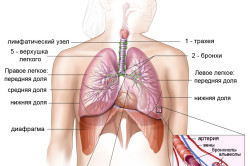
In the cutaneous lung, there is a top, bottom, three surfaces (rib, medial, diaphragmatic) and two edges (anterior and lower). On the costal surface of the upper part of the lungs, there is a furrow, similar to the clavicular artery, and in front of it there is a furrow of the brachial vein. On the costal surface, there is also a non-permanent mark of the 1st rib - subapical boron. The ribs and diaphragmatic surface of the legends are sharpened by sharpening the lower edges. When inhaling and vidihu, the lower edge of the legends is shifted from the vertical straight to the middle by 7-8 cm. The medial surface of the L. in front of it appears towards the costal surface, sharpening the front edge, and below the edge of the diaphragmatic surface.
At the front edge of the legends, there is a heart-shaped virality, which goes down to the tongue of the lung. on medial surfaces Both the leg and the backbone and the medical part, the heart are depressed. In addition, on the medial surface of the right legend, in front of the door, there is a depression from the adhering to the upper empty vein, and behind it, there are slight furrows from the adherence to the unpaired vein and the stravohodu. Approximately in the center of the medial surface of both legends, a funnel-shaped sinking is formed - the gate of legends. Through the gates of the lungs pass the head bronchus, legeny and bronchial arteries and veins, nerves gossip, lymphatic judgment; lymphatic universities are growing in the area of the head bronchial tubes. Pererakhovani anatomical statements in the present store the roots of the legends. The upper part of the lungs is occupied by the head bronchus, the lung artery and lymphatic universities, the bronchial nerves and the nerve gossip. bottom part winners take over the legends. The root of the lungs of the pleurora. At the bottom of the root of the legends, the duplication of the pleurisy pretends to be a triumphant Legendary sound.
Legends are stored in parts, adjoining one side of one interlobar gaps, which do not reach 1-2 cm root of the lung... The right legend has three parts: upper, middle and lower. The upper part is seen along the middle horizontal slit, the middle part of the lower part - as an oblique slit. In the legends of the two parts - the upper and the lower, divided by an oblique split. Portions of the lungs grow on the broncholegenic segments - the lungs, more and more isolated from the same suspension with full-tissue projections, in the skin where the segmental bronchus appears and appears on the other legendary artery; veins, draining a segment, bring in a roof at the veins, roztasovani in intersegmental partitions. According to the International Nomenclature (London, 1949), 10 bronchogenic segments are developed in the cutaneous lung. In the International Anatomical Nomenclature (PNA), the upper segment of the left legacy is from the rear (upper-posterior segment). The medial (heart) basal segment of the left leg is on the edge of the day.
In the skin segment, there are a few legeneous patches - those of the lungs, all the middle of which there is a reddening of the bronchus parietal (the second bronchus with a diameter of about 1 mm) right up to the end bronchiole; Particles emerged one from one and from one visceral pleura between lobular septa from fluffy fibrous with good fabrics... The cutaneous lung has nearly 800 patches. The growth of bronchial tubes (including the bronchial tubes) fix the bronchial tree, or the lungs.
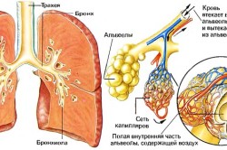
Dichotomous bronchial tubes dichotomously distribute to dichotomous (respiratory) bronchial tubes of the 1st to 4th order, which, in their own right, are subdivided into the alveolar ducts (walk), rasping from one to the point of developing, and ending with alveolar ducts. On the walls of the alveolar ducts, alveolar mice and dyshal bronchioles, they grow and appear in the holes of the alveoli of the legends. Alveoli at the same time with respiratory bronchial tubes, alveolar ducts and mincets fold the alveolar tree, or the dichal parenchyma of the legends; morphofunctional unit її acinus, which includes one dichal bronchiole and tied with it alveolar ducts, mice and alveoli.
Bronchioles in a single-ball cubic type of epithelium; they also contain secretaries and shirts. At the stage of endovian bronchioles, there are lamellas and cartilage plates. With the resulting tissue, the bronchial tubes drain off, go into the connective tissue basis of the dichal parenchyma of the lungs. In dichal bronchioles, cubic epithelial cells are absorbed; when passing into the alveolar ducts, the cubic epithelium changes into a single-ball flat alveolar epithelium. The alveoli wall, whisked by a single-ball flat alveolar epithelium, replaces three types of clitin: dichal (luskati) clitini, or alveolocyti of the 1st type, large (granular) clitini, or alveolocyti of the 2nd alveolar type, On the side of the epithelium, which is covered with a thin, non-lined ball, a surfactant - a speech, which is stored in phospholipids and proteins, and is violated by alveolocytes of the 2nd type. Surfactant volodya with kindly swirling surface-active powers, preventing the alveoli from falling to the vidiha, penetrating through the wall of microorganisms from the inhaled chew, overshooting the transudation of the day from the capillary. Alveolar epithelium grows on the basal membrane with a thickness of 0.05-0.1 microns. The call to the basal membrane adheres to the blood-bearing capillaries, to pass along the mjalveolar septa, as well as to the elastic fibers, to encircle the alveoli.
The top of the leg in the mid-stratum of the human appears to the dome of the pleurisy and protrudes through the upper aperture of the thoracic clavicle into the region of the sheath to the level of the top of the spinous ridge of the seventh spine ridge backward and 2-3 cm across the clavicle in front. The position of the cordons of the lungs and parietal pleura is similar. The front edge of the right legend is designed to the front chest wall on the line, how to carry out from the upper part of the lung to the medial tip of the clavicle, pushing up to the middle of the handle of the sternum and further downwards from the sternum line to the attachment of the VI costal cartilage to the sternum, to fix the lower border of the lung. The front edge of the left leg on the edge of the bottom of the IV rib from the sternum extends arcuately to the left and down to the point where the VI rib crosses from the peristernal line. The lower boundary of the right leg runs along the sternum line of the cartilage of the V rib, along the midclavicular line - the VI rib, along the anterior groin line - VII rib, on the scapula of the line - X rib, along the paravertebral line of the thoracic line - The lower boundary of the left legacy is seen from such a boundary of the right legacy, to repair the cartilage of the VI rib along the edge of the chest. In newborns, the upper part of the lung is located on the level of the first ribs, up to 20-25 years of stench reach the level normal for the growth of people. The lower boundary of L. new-born women are on one edge of a vishche, lower in the grown-ups, in the onset of fate they will go down. In people older than 60 years, the lower boundary of L. is 1-2 cm lower, lower in 30-40 people.
The ribs on the surface of the lungs stick to the parietal pleurora. With a whole back to the lungs, lie down between the ribs and the nerves, from them to the pleura and the intrathoracic fascia. Before the lungs lie on the open dome with diaphragms. The rights of the legacy to be seen as a diaphragm from the liver, to the left - from the spleen, the left nirka with the adrenal gland, the slunk, transverse colon and stoves. The media on the surface of the right leg is in front of the yogo it goes up to the right anterior, and the forehead - to the right brachiocephalic and upper empty veins, backward - to the stravohode. The media surface of the left legends є with the susids in front of the heart is drawn from the lynch, and the esophagus is through the arcuate aorta and the brachiocephalic vein, backwardly through the thoracic part of the aorta. Syntopy of the roots of the lungs is on the right and on the right. In front of the root of the right legacy, the viscid part of the aorta, the upper empty vein, the pericardium and the right anterior part of the heart; above and behind - an unpaired vein. Up to the root of the left legacy, the aortic arch lies above the top, and stravohid is behind. The offense of the root is in front of metinayut diaphragmatic, and behind - blukayuchy nerves.
Blood posture is associated with legionic and bronchial lysalosis with vessels. The legendary judges, who enter into a small blood circulation, are the head rank of the function of gas exchange. Bronchial judgments will make the lungs sick and give a great deal of blood circulation. Between two different systems, it is necessary to complete the bend of the anastomosis. The venous blood flows through the intralobular veins and flows into the veins of the interlobular septa. The veins of the p_dpleural tissue flow into the center. From the interlobular veins, intersegmental veins are formed, the veins of the segments are formed in a fraction, as in the gate of the lung, they are angry in the upper and lower veins.
With an ear of lymph lungs of the lung є the surface and the glyphs of the lymph capillaries. The superficial hem was welded into the visceral pleura. From her lymph go into the gossip of lymph judges of the 1st, 2nd and 3rd orders. Gliboka capillary hemisphere is located in the central tissue in the middle of the legenous patches, in the interlobular septa, in the mucous membrane of the bronchial tubes, near the intrapulmonary blood vessels and bronchial tubes. Regional lymphatic universities of the lung are equipped in the onset of the group: legenev, roztashovani in the parenchyma of the lungs, the leading rank in the misses of the bronchial tubes; bronchogenevia, which are covered in the area of hair growth of the head bronchial tubes; upper tracheobronchial, roseta on the lower part of the bi-surface of the trachea and in the tracheobronchial cuts; the lower tracheobronchial, or bifurcation, roseta on the lower surface of the trachea bifurcation and on the head bronchi; navkolotrachealnyh, roztasovany vdovzh trachea.
Innervation is good for legacy nerve gossip, as it is shaped by a bloating nerve, by the nodes of a pretty stovbur and a diaphragmatic nerve. At the gate, there is a lot of gossip going to the front and back. Х hіlki fix in the lung perіbronchial and perіvasal gossip, which supervise the redness of the bronchial tubes and blood-bearing vessels.
DOSLIGENNY METHODS
For the development of ailments of legends, vicorisation of illnesses and treatment of illnesses, as well as a number of special methods. Naybilsh characteristic scargs in case of sickness are legends є cough (dry or with phlegm), hemoptysis, sagging swelling, attack of breath, pain in the breasts, develop weakness, lustfulness. Anamnesis of ill health and life to follow the rules of the house. On'є, palpation, percussion and auscultation. Methods of self-improvement diagnostic value in case of legacy pathology and in the meaning of the world, start a large number of additional (laboratory, radiological, instrumental) dosages.
When looking at a sick animal, I especially respect for their position in the sled, the shape and symmetry of the breast, the character and the equitableness of the dysfunctional excursions, the camp of the mid-sized industries, the form breastfeeding of the ridge, frequency and depth of dichotomy, spіvіdnіnnіy phases і vidіkh, as well as on the preparation of worms and visible mucous membranes, the shape of fingernose phalanges (in wiggly drumsticks) and nіgtіv (in wiggly wiggles) clarify, the number of vibratory veins, liver enlargement, ascites, peripheral ridges.
Palpation of the chest wall gives the possibility of developing a zone of ailment, resistance, swelling, and especially crepitus during infantile emphysema, as well as arising the turn of the phenomenon of voice tremor.
For additional percussion, the boundaries of the lung are established, the rupture of the lower edges; from the changes of percussion sound to judge about the manifestation of pathological processes in the legends and pleural emptying.
Auscultation allows for the development of respiratory noises characteristic of the bronchogenic pathology of the disease, incl. wheezing, crepitations; viznachiti steps conduction to the voice of a sick person on the chest wall (bronchophonia). At normal, we have sounds, we are sick, we get off during auscultation like a dull sound; in case of infringement of the legacy tissue, bronchophonia will be felt, over the zone of atelectasis and pleural vipot vona weak.
Three special methods are most important for radiological preliminaries, including the order of ligamentous X-ray or large-frame fluorography, not less than in two projections, which are carried out after the bronchoscopic X-ray imaging, tomography. Everything is wider for the lightening of the lungs Computer tomography... For the follow-up of a small cola, blood circulation can be obtained from vicoristan angiopulmonography.
Of the instrumental endoscopic methods of advanced bronchoscopy, the most important is bronchoscopy; morphological dosage There is a special significance in diagnostics of poohlin from different localization. Otrimannya bronchoalveolar zmiyu before the hour of bronchoscopy and the last time may be very important in the diagnosis of bronchoalveolar disease. For additional thoracoscopy, it is possible to visually diagnose the parietal pleurisy and the surface of the lung, if necessary, take material for histological preliminaries. Mediastinoscopy, when through a small crack in the area of the jugular fossa in the middle, to introduce a special instrument - mediastinoscopy, allowing the anterior middles to be delayed. In addition, before the hour of mediastinoscopy, biopsies of the growth in front middle pathological statements, as well as nabotracheal, tracheobronchial (upper and lower) lymphatic universities, The camp of the bagatokh vipadkah (especially when evil new ideas) Imagining the nature and broadening of the pathological process in the lungs and bronchi.
Bіopsіya legenevoї tkanini i vnutrіlegochnoe roztashovanih patologіchnih utvoren Mauger zdіysnyuvatisya pid control rentgenotelevіzіynogo ekranu for dopomoga spetsіalnih gnuchkih іnstrumentіv (bіopsіynoї schiptsіv) scho conductive in legenevu tkaninu through stіnku bronchus at bronhoskopії (transbronchial bіopsіya) abo Shlyakhov punktsії through breast stіnku bіopsіynogo Golko rіznih konstruktsіy (transthoracic biopsia). In case of cases, if the method does not provide enough for morphological adolescence to a large amount of material, it is necessary to develop a biopsy of legacy tissue for intratracheal anesthesia through a small size of the breast; tse doslіdzhennya is most significant in the differential diagnosis of diseased diseases.
Functional methods of pre-examination allow assessing the anatomical and physiological power of the structural units of the lung and the adequacy of these processes, in order to prevent gas exchange between the children and the blood of the lung capillaries. Spirography gives you the ability to graphically rebuild the mental habits and to let you know the message in an hour. To add up to it, write down the speed of your lungs. On the whole principle, there are a large number of modern devices, which allow automatically calculating a number of indicators of legacy ventilation. when recording dikhalnykh rukhiv To reach the maximum amplitude of the snake obsyag legen with a quiet (life) and forced (forced life) dichanna. The emphasis on lung discharges in case of forced vidih is to visualize the support of the function, to be repaired by the ventilation apparatus as a whole, however, the main role in this type of visualization is to visualize the loss of prognosis dikhalnyh nobles... For a crooked, forced vidiha, you should get a quick vidiha in one second (OFV1), a maximum volume of fluidity to a stream (POS), the maximum volume of fluidity for a vidihu 25, 50 and 75% FZhEL50, MOS25, MOS FEV1 / VC - indicator (test) Tiffno.
Vvazhayut, the lowering of the maximum volumetric properties of the other half of the video (MOS50 and MOS75) early stages deterioration of transitory bronchial tubes, which can be victorious during screening diagnoses. In case of pathological processes, there is an expansion of the lung (pneumosclerosis, puffiness, pleural vipitis), the passages of the dysfunctional paths do not decrease, but the VC does not change. For a more accurate separation of obstructive and interjugal (restrictive) options for impaired ventilation, it is not very important to diagnose it, it is necessary to introduce the structure of the backward lungs, the maximum , OOL); The rest cannot be stopped when restoring the response curves. For vimiruvannya OOL vikoristovuyut barometric and convection methods. Up to the first, there is a retinal plethysmography, which allows for the presence of air-filled lungs, or, more precisely, for gas, to take place in the chest empty and upper dysfunctional paths, including and not ventilated areas (great bully). Convection methods of measuring OOL are based on the principle of changing and imaging from a light inert indicator gas in open and closed systems, and the values that have been obtained characterize only the ventilation of the air. Obstructive impairment of ventilation can be prevented by yak with little change, as well as with a change in VC. The first one will see an increase in the OEL and the general increase in the OOL, and in the other, the OEL becomes normal, and the OOL builds up.
Zagalny plethysmography also allows for direct characterization of the bronchial support in the minds of the spokyny dichannya (Raw). Behind the shape of the loops, it is possible to visualize the depletion between the flow of water and the grip of the middle chamber to the apparatus, to help the sick person, there can be clear indications of the presence of foul-ventilated zones of light and inhomogeneity;
For an unprecedented characterization of the elastic powers of the legends, one must immediately write down the transpulmonary vice, as a way of reestablishing the internal vice, and dysfunctional communication in static Lung tension (GI) is reduced to a single transpulmonary vise at a given curve. With pneumosclerosis, GL changes, and with emphysemia, it grows.
The diffusional building of light for carbon oxide (DLSO), which comes close to the diffusional powers to the sourness, changes when changing the environment on the level of OEL (DLZD), or in the steady state (DLus). Otrimuvani indicators represent the integral characteristics of the minds of gas exchange in the legends, the odds of lying not only because of the diffusional powers of the alveolo membrane, but because of the irregularity of the minds of ventilation, and also because of the factors. The value of DLz is to be found in the leading rank from the functional surface of the legends, and the DLus is to be found in a larger world because of the equal value of the regular ventilation-perfusion signals, but with one-hour monitoring of the dosage methods
efficiency ventilation legends assess according to the dynamics of the physiological dead space to the dical physical options). Retardation of alveolar-arterial changes in acidity in case of post-inhalation normal, hyper- and hypoxic sums, as well as in the development of the mechanism of loss of gas exchange (manifestation of arteriovenous diarrhea)
The results of the efficiency of the system and the outward reaction characterize the sourness in the plasma of arterial blood (pO2) and the increase in hemoglobin sourness, as they visualize the total statuses of all processes, which will prevent the oxygenation of the blood. The exchange of two oxides in coal is characterized by a partial pressure in the plasma of arterial blood (pCO2), as well as in the acidic pool of blood, in a direct world of adequacy of ventilation. For the value of рСО2, the microanalyzer of Astrup is used to determine the value of pCO2; рО2 can be used for additional attachments to Astrup microanalyzers or a special attachment. Oximetry should be used for the vivification of blood-sour blood.
In case of pre-existing bronchial passages due to the appearance of adherent bronchospasm and the appearance of bronchial reactivity in the presence of pharmacological tests with the ingestion of drugs, as well as the development of weakness of the bronchial muscle spasm.
For the development of regional lung functions (ventilation, blood flow) the most effective radionuclide methods. In order to assess the regional ventilation of vikoristovuyut inhalation 133Xe, for the assessment of the regional blood flow, intravenously inject the microaggregates albumin, micheni 131I or 99mTc; then to conduct a radiometer or a radioisotope scan of the lung behind additional auxiliary attachments (for example, a gamma camera), automatically calculating a number of functional indicators. Tetrapolar rheopulmonography is less opportunities for the implantation of the regional blood flow in the lungs - to increase the electrical support of the lungs, to lay down all the blood supply.
Regular ventilation is also followed by additional X-ray functional methods, based on the change in the progression of young lungs in the phase of the dyshal cycle. The simplest of them is the tomorespiratory test: the value of the penetration of the leg tissue according to the tomograms, the morbidity of the vidiha and the vidih. We will thoroughly use the X-ray functional method, allowing for sufficient accuracy in terms of the significance of the regional ventilation changes, pneumopolygraphy, with such signs of the lungs in the inhalation phase and in the form of a healthy state of health.
I have a very important role in assessing the legacy of blood flow in the case of diseases of the legends of blood circulation and, in the first place, making a grip in the legacy of arteries to clarify the level of legacy hypertension. Nepryamі Metodi doslіdzhennya legenevogo blood flow (by rentgenogramah, elektrokardіograma, kіnetokardіogrammam viyavilisya nedostatno accurately. Digit bіlshu vіrogіdnіst vimіrіv vise in legenevіy arterії i row pokaznikіv right shlunochka i gemodinamіki small stake krovoobіgu zabezpechuyut ehokardіografіchnі i Doppler kardіografіchnі method. For Relief direct zonduvannya legenevoї arterії can accurately vimіryat the grip in nіy and count a number of hemodynamic indicators (for example, the back of the legacy of the judiciary, to the robot of the right dumbbell).
With all the legends fall ill, carry out laboratory dosage, Zokrema analysis of blood and sech. Special meaning of the analysis of the harkotinnya. So, її bacteriological progress is given the opportunity to establish the etiology of the infectious process in the legends. The pre-treatment of a cellulite storage of sputum in a row of diseases (for example, in case of bronchogenic cancer) allows you to clarify the diagnosis. Bacteriologic and cytologic dosage of pleural exudate additionally helps in determining the etiology and nature of pleurisy, which accelerates the legacy of discomfort. Great value is given to the material that is not obstructed by the microflora of the upper dichally nobility; It is necessary to remove it from the trachea, bronchial tubes and alveoli (smears and bronchoalveolar serum during bronchoscopy, aspirate when puncturing the trachea), as well as from infectious disease in the legends. Material for virologic dosage (immunofluorescent method, cultivation of viruses) є scrambling of the mucous membrane of the nasopharynx and tracheobronchial tree. To clarify the ethiological factor of bacteriological and virological preliminaries, add serological (the value of titers of antibodies to bacteria and viruses). Biochemistry of blood pressure (proteinogram, the value of C-reactive blood cells, sialic acids, haptoglobin) is carried out with the help of determining the activity of the bronchogenic bronchogenic process, which is functionally important for vital organs, especially for the liver. in case of a decline in the amount of legends). Immunological advancement is possible to assess the specificity of the reactivity of the sick person, the quest for the effectiveness of the treatment and the establishment of an indication before the immune response.
PATHOLOGY
Pathology of the leg includes developmental defects; pneumopathies of the new genital; recessionally amazed; ushkojennya; sickness, ethiologically tied with biological pathogenic animals; enfeebled, zumovlenі injected shkіdlivny chemіchnіh і physical factors; chronic nonspecific ailments; becoming ill, pathogenetically tied with allergies; desemination of illness; pathological conditions, tied with damage to the lung blood circulation.
Flaws in the development. Prior to the most frequent defects in the development of the lungs, tied with incompletely damaged anatomical structural and tissue elements, there is agenesis, aplasia, hapoplasia and the growth of localized emphysema of the legends; up to the flaws, which are characterized by the appearance of superfluous disembriogenetic establishment, - a solitary lung (part, a segment) with an extraordinary blood loss, an additional lung with abnormal blood loss (sequestration), Wad the development of Sudin legends of key importance may arteriovenous fistula. ...
Agenesis and aplasia. Under the agenesis of the lungs there is a visibility of the lung and head bronchus, under aplasia - the visibility of the lung or part of the presence of a formed abnormal bronchus. Agenesis of vines as a result of the growth of bronchogenic nirok at the 4th stage of the intrauterine life, aplasia - at the time of the start of the development at the 5th stage.
With bilateral agenesis and aplasia of the lungs, children are not published. The clinical picture of one-sided agenesis and aplasia of the lungs is similar and characterized by dysfunctional asymmetry (due to the changes in the act of affecting the affected side of the chest wall), dulling of the percussion sound, as well as dulling the increase in intensity. Clinically and radiologically, symptoms begin to change the middle of the battle. On the x-ray of the chest wall, there can be a total shading of half of the breast emptying, and in an hour you can move a part of the healthy lung to the opposite side (a symptom of mediastinal hernia). At the same time, there are clinical and radiological signs that are very similar to the symptoms of atelectasis of the legeny in newborns, to clarify the diagnosis of vikoristovy, bronchoscopy, bronchography, angiopulmonography. Operative treatment of agenesis and aplasia of legends, as a rule, is not possible. Forecast for life with a one-sided development of hospitality.
Hypoplasia is an incomplete condition of all structural elements of the lungs of any part (part, segment). They develop two of the most widespread forms of hypoplasia of legends - simple and cystic. A simple hypoplasia is characterized by an equal change in the lungs obscuration of any part, in the sound of the bronchial tubes and in the diameter of the lungs. It is a clear picture to lie down in the course of the battle and the manifestation of either the presence of the ignition changes in the hypoplastic or the sum of the lungs with it. There may be signs of dysfunctional deficiencies, asymmetry of the breast and dysfunctional asymmetry, clinical and radiological symptoms of the change of organs in the middle towards the change in the general population. In case of impaired lung ventilation, secretory and drainage function of the bronchi, there may be such signs as dullness of the percussion sound and weakening of the dullness, dry and vologic pulmonary wheezing of tissue. To reach the often hypoplastic part of the legends, a gn_yno-ignition process develops, which will add a key picture to the main picture. Repeated ignition processes in the singing of the legends є by the drive in order to detect the hypoplasia of the legends. Bronchoscopy, bronchography, angiopulmonography, radionuclide scan of the lungs are allowed in the cycle of bronchoscopy, bronchography, angiopulmonography, as a rule, clarify the diagnosis. During bronchoscopy, there are stages and localization of ignition changes, options for the detection of bronchial tubes and stages of sounding of the mouth. On the bronchogram, there is a change in the general lung, as a rule, deformations of the bronchial tree. On an angiopulmonogram, the value of blood flow can be determined. Radionuclide methods of latency allow to set up steps of ventilation and blood flow in the zone of defect development. It is shown before the operative correction to lie down in the steps of functional breakdowns and variations in clinical manifestations. Promptly lіkuvannya polyagaє often in the seen underperforming іddіlіv legends. The operation can be a Viconan in any occasion. Forecast to be the head of the rank as a result of the obsyagu battle and also on the basis of manifestation of for operational acceleration.
Cystic hipoplasia (congenital polycystosis of legeny) is a defect of development, with any terminal appearance of the bronchial tree on the equal subsegmental bronchial tubes or bronchial tubes may cause cystic enlargement of the expanding diameters. Clinically, cystic hypoplasia is a little less likely to occur as a result of downtime. On the roentgenogram in the battle zone, there can be many subtleties of empty empty spaces, as a rule, not to revenge the age. The triviality of finding such empty spaces, accumulating a secret in them, their consumption and information is superfluous, as a rule, with a key picture. ignition process in the lungs. At the same time, the most characteristic signs of intoxication, vologic cough with gnaric sputum, symptoms of dysheal deficiency. Radiologically, in the whole period, there can be many rіvnі rіdini in cystic emptyings.
With a trivial flow ignition process, it is not easy to find difficult in the differential diagnosis of cystic hypoplasia of lesions and bronchiectasis. In some cases, cystic hypoplasia is often considered a disease of fibrous-cavernous tuberculosis, and such ailments were trivial and unsuccessful to take anti-tuberculous drugs. Retaliation assessment of anamnestic tributes, clinical and radiological pictures, as well as the results of special methods in advance, allow in a large number of cases to establish a diagnosis before the operation. For the prevention of tuberculosis of the lungs, carry out bacteriological preliminaries of sputum, tuberculous tests, immunologic preliminaries.
Lіkuvannya operatively і polyagaє at the seen urazhenoja part of the legends. Before the operation, it is possible to reduce the ignition process as much as possible, allowing the reduction of hundreds of operational acceleration and improvement of the results of the operational treatment.
When pіdtverdzhennі Just abo kіstoznoї gіpoplazії legenіv (on pіdstavі rezultatіv morfometrichnogo doslіdzhennya vіddalenіy chastinі legenіv) neobhіdno postіyne dispensary sposterezhennya for the ailing, to scho no mozhna viklyuchiti nayavnіst have less then virazhenih torn down in Structural Elements, scho zalishilisya vіddіlіv legen, scho Mauger lead to rozvitku in them fiery zm_n.
Vrodzhena localized emphyzema (vrodzhena chastkovaya emfizema, hypertrophic emphysema) is a defect in development, which is characterized by stretching of parenchyma parts of legends (as a rule, one part). Deyakі authoritarian pov'yazuyut Yogo viniknennya of aplazієyu hryaschovih elementіv bronhіv, gіpoplazієyu elastichnih fibers smooth m'yazіv kіntsevih i dihalnih bronhіol i іnshimi torn down in the structural odinitsyah legenevoї tkanini scho stvoryuє peredumovi for viniknennya valve mehanіzmu scho spriyaє nadmіrnomu zduttya vіdpovіdnoї Chastain legenіv.
The clinical picture is characterized by the syndromes of dichotomous and heart-vascular insufficiency, the swelling of some of them can be quite different. The visualization is decompensated, subcompensated and compensated for the localized localized emphysema of the legends. With decompensation of congenital localized emphizemia cleverly show find out all at once from the people. Most often, cyanosis, dysfunctional, dyshally asymmetry, uncomfortable, frequent dry cough, attack asphyxia before an hour of the year are often spontaneous. Virіshalny in diagnostics є radiologic diagnostics. On the roentgenogram, there may be an increase in the progression of the leg tissue, as much as an additional increase in the appearance of the lung malignant, the change in the middle (one symptom of mediastinal grizzly), the collapse (slowing down) of healthy legends. The appearance of the remaining signs is extremely important for differential diagnostics s pneumothorax.
With subcompensated congenital localized emphysema of the legends, the symptoms of a lesser turn are described, and they become more thoughtful in children of the first fate of life when they are restless, and in a larger older person - when they are physically fit.
When compensating for the built-in localized emphysemia of the classical legends, they can manifest themselves in the edge with weak turns, unacceptable. It is not often the case that the ignition changes in the affected lungs are not collapsed є by the drive for the X-ray dosage, as it allows the characteristic snakes in the legends to emerge. Some of the signs of localized emphysemia of legends appear during angiopulmonography (with decompensation of the form, it is indicated through the severe camp of the sick person): in the zone of advanced progression, the legends appear to the least extent of the lungs, the cases of judgment are severe. Radionuclide before long-term blood flow has been shown to be a significant decrease in all types of diseases.
The only method of detecting an innate localized emphizemia of legends is an operative one (a visible part of the lesion). The operation can be a Viconan in any occasion. The forecast is to be the head of the rank of obsyagu urazhennya.
Dodatkovo lung (part, segment) with extraordinary blood circulation can be normally formed and functionally povnotsinny. Such a developmental defect is not less of a key value and appears vaguely in radiological dosage. However, part of the structural elements of the pre-donated part of the segment of the lesions is incomplete (hypoplastic in the pre-pulmonary). In many cases, clinical tactics are shown as well as in case of hypoplasia.
Sequestration is a defect in development, when it is hypoplastic, it does not come from the bronchial tree of the main lung part; Venous blood is from such a dilyanka, as a rule, it is introduced into the system of a small blood circulation, or, more quickly, into the system of an upper empty vein. A part of the lungs is hypoplastic due to abnormal blood loss, it can be seen in a single bone, or polycystic illumination, a posture of the lung tissue of the main lung, and a small amount of pleural tissue (extrapulmonary sequestration), or in the middle of the lung (intrapulmonary tissue) of the main lung. Most often, sequestration is spontaneous in the lower medical records of the legend. In the literature about the localization of the sequestration of the lungs in the black empty.
Klіnіchnі show up in childish in case of information and initiation of the ignition process in the affected and adjoining normal views of the leg. Before them, the loss of self-esteem, temperature adjustment, as well as physical data, characteristic of frequent pneumonia, are referred to. The presence of quiet symptoms is not only due to the stages of ignition changes, but due to the nature of the hypoplasia (simple or cystic), and also due to the localization (postgenevia or intrapulmonary) of a vicious delay.
Diagnosis of lung sequestration is important. With intrapulmonary sequestration on an oglyadovaya X-ray of the thoracic clitine, it is possible to start a slowing down of the lung tissue of a sizeable volume, similar to that in case of pneumonic infiltration. If an abnormal judgment is detected during aortography, or during tomography, it will be possible to make a diagnosis before surgery. Lіkuvannya operatively - vividly urazhenoї dіlyanka legen. The prognosis is hospitable and will remain the head rank during the interruption of the operational period.
Vrodzhena solitarna kista - cystosne illumination, roztashovane centrally, so in the rooted zone, or closer to the periphery of the legacy. In lіteraturі zustrіchayutsya th INSHI titles tsogo vice rozvitku: bronchogenic kіsta, Bronchial kіsta, that scho at mіkroskopіchnomu doslіdzhennі stіnok kіstoznih utvoren them in bіlshostі vipadkіv viyavlyayut Elements bronhіalnih stіnok - plate hryaschovі, tsilіndrichny epіtelіy, elastichnі, m'yazovі i іn fiber. The emergence of congenital solitary cysts, mabut, is tied to the prescription of hypoplastic parts (segment, sub-segment) of the lungs, more often it is seen from the bronchial tree, because of the reason for it.
In case of small cysts, which does not result from the bronchial tree, it is usually possible to manifest itself as a daily and unremarkable radiological sign. When the brush is broken from the bronchial tree, there may be symptoms, which are accompanied by partial drainage in the place of the brush through the bronchial tree: vologic cough, dry wheezing during auscultation. In case of infection of the brush, there may be symptoms of inflammation and intoxication (increased temperature, restlessness, decreased appetite, etc.). Great, centrally grown solitary brushes are often associated with a bronchial tree. The stench can stifle the meaning of the food and the legacy and lead to the development of a dysfunctional deficiency. The dysfunctional and heart-vascular deficiency can be explained by the changes in the valve mechanism in the cyst.
The peculiarities of the physical tributes are due to the size of the brush, the character and the lassitude. So, for the great and struggling men of the cysts, it is more characteristic of the weakening of the spirit at the battle, the sound of the box-type sound, the change of the middle to the opposite side (at the time of the winter of the middle of the battle) Kisti, memorized with a vivid vmistom (see if the stench may cause significant changes), rarely show symptoms, powering the exertion of the cysts; characteristic for them physical signs є weakening of the sound and dullness of the percussion sound on the side of the strike.
The inconvenience of recognition is stored in the place of a cyst of chickens and forecasts of overshoot (increase, suppression, breeding) є provided for prompt treatment. Most often there are polygons near the visible brush or the lungs. (Segment, parts) at once with a brush. Prognosis
Arteriovenous fistulas - pathological results of the formation of legeneous arteries and veins - leading to visceral forms of angiodysplasia, amalgamated with damaged development court system lungs at the early stages of the embryonic development. Localization of fistulas of the disease; more often the stench grows in parenchyma legends.
Klinichny show to lie due to the size, localization and nature of the fistulas. In case of the appearance of some great judges, the first plan is to develop hemodynamic changes, which manifest themselves as cyanosis, weakness, weakness, congestion, and some hemoptysis. Chronic hypoxemia is supressed by compensatory polycythemia and polyglobuli, lesions of the larynx of blood, as well as the diagnosis of legeneous bleeding. Can be removed in growth and physical development as a result of chronic hypoxia. Inodi above the lung, a judgment noise is heard.
The radiological picture is to be found in the form of changes. best characteristic symptomє the appearance in the legacy of fabrics of the shading of the small changes, the form and intensity. For additional angiopulmonography, localization of fistulas and shunt steps can be established.
Lіkuvannya operatively - resection of urazhenoi dіlyanka legen. The prognosis is to lie in the head rank as a result of the obsyagu urazhennya, as well as on the basis of the manifestation of the development of sudin in other organs.
Pneumopathies of neonatal infants include atelectasis of the legs, hyaline membrane of the ailment and embryonic hemorrhagic syndrome, surfactant deficiency. The stench develops more often in premature and immature full-term children in the first year of life (div. Distress syndrome of respiratory neonatal).
Spadkovo amazed. Naybіlshe values Sered them toil legenevі Show mukovіstsidozu and takozh spadkovy defіtsit іngіbіtorіv proteases in the main (a1-antitrypsin. When nestachі a1-antitrypsin vіdbuvaєtsya ruynuvannya naytonshih structures legenevoї tkanini nakopichuyutsya in nadmіrnіy kіlkostі proteases leukocyte, macrophage, pankreatichnogo i bakterіalnogo pohodzhennya. Zahvoryuvannya uspadkovuєtsya by autosomal recessive type. more often, the level of the protease inhibitor becomes 75-50% of the norm, except that you do not lead to the development of the developed emphysemia, ale, mabut, which is less significant in the pathogenesis of a number of chronic ailments L. . Described as a therapy for natural inhibitors of proteases (counterkal, gordox), inhibitors of the calcarein-kinin system (parmidin), as well as androgens. The prognosis of homozygous forms of ailment, as a rule, is unpleasant.
Possibility of the lungs to move at close and on display. Zakritі pozhkojennya turn on zabіy, zakryryv, hush and strus legends. At blows of the lungs of the vine intrapulmonary hemorrhage. When we cut through the legacy fabric, we go with the edge of the rib. Post-surgery of the chest wall can zoom in hemothorax, and post-surgery of the lung tissue - pneumothorax. A blow to the lungs reveals pain in the breasts, coughing up hemoptysis, and if the lungs are closed, there may be signs of a pneumocystis, hemothorax, pneumothorax. X-ray in the impact zone can be infiltratively darkened, in some part of the fall, light, gas і ridin in the pleural emptying.
Likuvannya polyagaє in usunenny pain syndrome(Alcohol-novocaine blockade of the area of rib fractures), aspirations of blood and blood from the pleural emptying by way of pleural puncture. When blood is purchased in the bronchial tree, the bronchoscopic period is lowered for an hour. It is more important to come in, direct to the problem of atelectasis of the lungs and pneumonia.
Disease of the legends of the winery as a result of a sharp intense squeezing of the breast tissue, often in a sagittal direct with, as a rule, spasmodic vocal cords; it is not uncommon to suffer from multiple bilateral fractures of the ribs. When the legends are pressed, it is indicated that the rapid expansion of the intrapulmonary vise, many alveoli, intrapulmonary hemorrhages, interstitial ridge are indicated. The winery of the gostra is dyshally lacking in the development of the "shock legacy" and the failure of ventilation through the ruptured frame of the chest wall. When large bronchial tubes are discharged, hemothorax and middle emphysema develop, which increases the rate of ventilation. As a result of raptus diagnostics of venous hypertension, there can be a lot of internal blood vessels, which can produce shkіrі, especially on the face and upper part of the tulub, Cyanotic filling.
Lіkuvannya includes kisnevu therapy, sanation of the bronchial tree. In case of uncorrectable hypoxemia and hypercapacia, it is necessary piece ventilation lungs with a positive grip in the end of the vidih and in the go, straightened on the weakening of the respiratory distress syndrome.
The result is a penetrating stabbing or bursting injury to the breasts. Disruption of vital functions with injured lungs results in traumatic pneumothorax, hemothorax, hemorrhage, as well as blood loss in the mental paths and obstruction of the rest, which can lead to a severe mental deficiency in late Signs of the ear of legends in case of wounds of the breasts є hemoptysis, coughing up gas through the wound, infantile emphysema in the circumference, pain in the breasts with stiffness, backwardness and dysfunctional deficiency. Physically, there may be signs of pneumo- and hemothorax, which can be confirmed radiologically. For the additional radiological dosage in the lung, it is possible to see a side body (in case of injury), in the soft tissues of the chest wall - gas projection.
The first help polyagaє in an applied dressing (in case of an abnormal valve pneumothorax, she is to blame for her sealing), giving the patient a seated position, acid therapy. The treatment is carried out in a hospital and includes entering, directing to the liquidation of pneumo- and hemothorax, after the direction of the damaged lung and blood loss. With mild cases without hemothorax and pneumothorax, it may be symptomatic. With an insignificant spontaneous sealing of the ear lesions with a small pneumothorax and (or) hemothorax, a pleural puncture will be needed to evacuate blood and nutrition. With larger important ears and leakage of the leg tissue, and the pleural emptying, drain actively with a tube (internal diameter not less than 1 cm) at the eighth intercostal space along the posterior groin lines and additional systemic support for other In the case of a large number of vapors, I will prevent the expansion of the lungs with a stretch of 1-3 dB. It is not often shown before the operative lykuvannya. They are a great defect of the thoracic wall, which is a vimag of an operative closure (pneumothorax induction); bleeding trivially in the pleural empty space, or in the dizzy way; the inability to open a vacuum during active aspiration instead of a pleural emptying for 2-3 dB; non-stop stress pneumothorax; the formation of a straining blood clot in the pleural empty space ("flared hemothorax"), missed fibrinolytic drugs; great side body. The entitlement includes surgical treatment and suturing of chest wounds, thoracotomy, hemostasis, suturing of lung wounds. At the time of the great roztroshchennya legacy tissue, one should carry out an atypical resection of the leg, in rare cases - forehead pneumonectomy. Most frequent wounds of the lesions the rate of pleurisy, bronchial fistulas, which usually occur, as a rule, when ill-feeling are unpleasant, promptly distribute the legends and liquidate too much empty, as well as pneumonia. The forecast for a large number of vapors is hospitable. Lethality in peacetime does not change 2-4%.
Poisoning, ethiologically linked with biological pathogenic pathogens (bacteria, viruses, fungi, most common, helminths). The greatest significance of the disease of the group is pneumonia, as well as abscess and gangrene of the leg.
Abscess and gangrene of the lung represent the state of infection and destruction of legends. In an abscess, they easily knock out a larger amount of empty material, as it is formed as a result of the fiery melting of the legacy fabric. Gangrene is characterized by a great, but not subtle to intermittent necrosis and rotten tissue growth.
Inducers of abscess and gangrene, in the first place, non-spore-forming anaerobic microorganisms (bacteria, fusobacteria, anaerobic coca and in.) The pathogens should be treated in the Legenev tissue, often transbronchial, before hematogenous (for example, with sepsis). An important factor in the development of cyx infections and the formation of a destructive process, reduction of muscle and backward reactivity as a result of viral and bacterial (pneumonia) infections. In a large number of cases of development of abscess and gangrene, the legacy is associated with the aspirations of the information material from the mouth emptying, which is facilitated in case of decreased cough reflex (for example, in case of alcoholic, cerebral disease). Aspіratsіyny mehanіzm characteristic for іnfektsіynih destruktsіy anaerobnoї etіologії scho pov'yazano of great kіlkіstyu neklostridіalnih anaerobіv in rotovіy porozhninі, Especially when karієsі zubіv parodontozі i, i viniknennyam at aspіratsії bezpovіtryanih dіlyanok legenevoї tkanini (atelectasis) in yakih stvoryuyutsya spriyatlivі minds for rozmnozhennya anaerobnoї mіkroflori ... In addition, abscess formation occurs on the side of the bronchial tubes, as well as chronic illnesses ( diabet, chronic obstructive bronchitis, get sick blood-forming organs), Trivale stasis of immunosuppressors. With hematogenous inflammation, there is an embolism of the hematogenous artery disease.
Abscess and gangrene of the lungs are often observed in people of the middle age, especially in alcohol. The abscess is easy to fix, as a rule, it is gross - s nezduzhannya, chills, fever, chest pain. Before I burst into the bronchial tree, I started to get rid of the cough from the daytime or insignificant. Physical signs indicate massive (angry, private) pneumonia. Characteristics of bends of leukocytosis from a leukocyte formula in the presence of a leukocyte formula, increased by SHOE. Radiologically, in the cob period, the disease begins to be massively obscured by the leg tissue, and pneumonia is interpreted.
At the time of the breakthrough of the gnarium in the bronchial tree, the tree is key picture Adequacy of emptying and fast melting and processing of necrotic substrate begins. In the rapid natural drainage of ailments, there is a great number of rotten diseases, often with an unacceptable rotten smell, phlegm, the temperature and symptoms of intoxication change, it is not possible to catch up on the background of the information. Nadal іnfіltratsіya change, the level of birth, and the empty itself deforms, changes. In 1-3 ms. You can come outside of thinning empty empty spaces, for it is so called dry thinning empty empty without critical manifestations.
With a filthy drainage emptying and (abo) the hopeful melting of a necrotic substrate of ailments, it takes a trivial hour to cough up phlegm, a feverish woman with chills and sweats grows, it takes a trivial hour to cough up phlegm, a fever with chills and sweats, grows intoxication. The number of disclosing a sick old is earthy-zhovtim, the finger phalanges of fingers are deformed, with a whole finger they swell the eyes of drum sticks, nigti - year old glasses. Anemia, hypoproteinemia grow, there is a block in the battle. Radiologically, there is a build-up of infiltration of legacy tissue, and a level of age in the empty space (empty) occurs.
Clinically gangrene of the lungs is unpleasantly streaming abscess of legends, ale it turns out to be even more vague. When you appear ryasnoi (up to 500 ml for doba), pick up stinky sputum, when dispensed into 3 balls, do not bring the sick person lodging. Radiographically, the ear shows the sputum on the last of the great lingering, as a rule, it borrows 1-2 parts, or everything is easier, if the wrong form starts, it’s a lot of middles of education, some of them in the rіdynia. Symptoms of intoxication quickly progress, often there is a dizzying lack.
In case of gangrenous abscess of the legends of the class, show a little less turn, less with gangrene L. great razmіrіv, Wrong internal contours (attached sequestration) and incorrect delinks obscuring the middle of it (vilny sequestry). Around empty it is a trivial hour to save a lot of information, like when friendly perebіgu change every now and then.
Unpleasantly flowing abscess, gangrene and gangrenous abscess of the lungs can be delayed by pneumothorax (pus suppurating and if I get rid of the empty pleural space in the lungs), pulmonary hemorrhage Qih vipadkah has a very lethal result.
Diagnosis grounded on characteristic clinical and radiological signs. For the recognition of ethiotropic treatment, it is necessary to establish ethological factor... At the same time, it is necessary to carry out bacteriological preliminaries (seeding) of the material, cut off by the way of the point from the drop (infiltration), pleural emptying, trachea. Wetness until lidzhuvati is not recommended in connection with the manifestation of the microflora of the upper dichally nobles. Cultivation of microorganisms is bazhano grow according to aerobic and strictly anaerobic methods. In case of unpleasantness, an anaerobic microflora can be carried out by means of metabolites by the method of gas chromatography with pus. Anaerobic nature of the process can be made by rulings and deeds key signs(A case for aspirations in the anamnesis, stinky odor and siruvate colorectal sputum and pleural effusion, strength to the process until it expands on the chest wall with punctures and draining due to anxiety disorders)
Differential diagnosis is carried out, in the first place, with destructive forms of tuberculosis of the lung, festering cyst of the lungs, and also with cancer of the lungs. For destructive forms of tuberculosis, it is a leg. characteristically mensheed intoxication, torpid overrun; in the sputum, there is a mycobacterium tuberculosis. With a festering cyst of the lungs, the intoxication is rotated insignificantly, near the thin emptying of the day, the infiltration is rotated. When the cancer of the legends falls, the sputum is wretched, odorless, intoxicating and feverish; emptying of materials and an uneven internal contour; Diagnosis is confirmed by the presence of sputum (appearance of puffy clitins) and biopsies.
Mild abscess and gangrene treatment is predominantly more conservative in the presence of active surgical and endoscopic manipulations. It includes three binding components: optimal drainage of empty empty spaces and active sanitation; suppression of pathogenic microfloria; renewal of existing reactions to the organism of ailing and damaged homeostasis. To ensure optimal drainage of empty spills, vikoristovuyut from hardened bacteria, bronchial infections, mucolytics, proteolytic enzymes, postural drainage. Repeated bronchoscopy with catheterization and drainage of bronchial tubes is more effective. Under study is kateterizatsіya traheї i drenuyuchih bronhіv of administration bronholіtikіv, mucolytics, antibakterіalnih zasobіv i aspіratsієyu harkotinnya Mauger zdіysnyuvatisya through thin drainage tube inserted into the trachea Shlyakhov її punktsії (Mіkrotraheostomіya) Velikі subpleurally porozhnini mozhna sanuvati Shlyakhov transthoracic punktsіy abo mіkrodrenіrovanіya of promivannyam antiseptichnimi rozchinami i Introduction antibacterial diseases.
The pathogenic microflora is mainly driven by antibiotics, which are administered, as a rule, into the upper empty vein through a special catheter. When aerobic microflora is seen, synthetic penicilli, as well as antibiotics, are shown wide range diy, especially cephalosporin (for example, cefazolin); anaerobic - great doses of penicilin, levomycetin, metronidazole (trichopol).
Come on, according to the renewal of the power of the ailing organism, to include a retreat, high-calorie consumption, a lot of vitamins, repeated infusion of blood preparations, as well as the development of electrolytes for the solution of water-soluble substances. For the stimulation of immunological reactivity, immunocorrecting drugs (sodium nucleinate, thymalin, levamizol, tactical and in.), UV-optimized blood. In severe forms of illness, hemosorption and plasmapheresis are indicated. Operative treatment (resection of legends or pneumonectomy) is indicated for non-efficacy of the conservative politics, And also in a large number of cases with extended gangrene L. as one of the sick people; it is carried out for the maximum possible compensation of homeostatic damage.
The prognosis for abscesses and gangrene is light, as a rule, serious. The lethality with abscesses of L. reaches 5-7%, and with extended gangrene, the lungs are up to 40% or more. In 15-20% of cases of lung asbestosis. move to chronic form When a colic gnarium is formed on the mallet, it is formed incorrectly to form empty, whipped by granulations, with fibrous changes near and periodic sharpening of the infectious process. The main method for the treatment of chronic abscesses is the operative one: the visualization of the affected part of the abo (more) the lesser part of the lungs, one of the whole lung.
Specific disease of bacterial nature. We will expand on them є tuberculosis of the legends. The syphilis of the lungs in the wicked minds grows in the edge. With natural syphilis lesions, diffuse depletion, fibrosis of the interstitial tissue, abnormal development of the alveoli, the appearance of cubic cells, the manifestation of bladder treponema in the alveoli is indicated. To be seen among the dead people, or among the new people, as they go in the first days of life. Nabutia syphilis of the lungs. Sposter in the tertiary period of ailment and is characterized by the development of the gum of the lungs and (more) diffuse lung fibrosis. Diagnosis should be put on the diagnosis in case of x-ray preliminaries in the legends and positive serological reactions to syphilis. In order to confirm the diagnosis, carry out a biopsy. Lіkuvannya also, like і with other forms of tertiary syphilis.
Fungal disease. The fungal flora can be the reason for a number of chronic diseases of the disease. - pneumomycosis.
Zahvoryuvannya, scho viklikayutsya simplest. In case of amoebiasis, which is called Entamoeba histolytica, in most cases, the intestine is primarily affected, then liver abscesses form. It is easy to get sick to a pathological process when the amoebaea abscess develops through the diaphragm. Early amoebic abscess of the liver disease is hematogenous without liver damage. The ailment to scars on pain in the breasts and cough with clear brown phlegm, in case of microscopic dyslips, there may be amoeba. Rohigh standing of the right dome with diaphragms, empty with a horizontal line of the line, sagging in the lower viddylah of the lungs. Likuvannya is also, as in the case of other forms of amoebiasis, in some cases it is necessary to microprocess an empty abscess or to accelerate pleurisy.
In case of toxoplasmosis, where Toxoplasma gondii occurs, granulomas can form in the lungs with occasional necrosis, we will drain lymphocytes and plasma cells; granulomas are slender until they are squeezed. In case of lesions on aphids, showing toxoplasmosis, there is a cough and wheezing. Radiologically, there are many other types of flares in legends, some of them. Laboratory diagnostics and lіkuvannya also, yak at іnіh forms of toxoplasmosis.
Pneumocystosis, accumulation of Pneumocystis carinii, develop as a head rank in case of impaired immunity, incl. with a syndrome of an accumulated immune deficiency (div. VIL-infection).
Zahvoryuvannya, scho viklikayuyutsya helminths. The most significant is the average of small echinococosis legends, Echinococcus granulosus mummations. Vin is characterized by the development of the brush, as it usually does not manifest itself clinically, and it can be detected vividly during radiological diagnosis. In the world of increased brush size and crushed tissue, there are pains in the breasts, cough (sometimes dry, with wetness, sometimes bloody blood), backside. With large cysts, the deformation of the breast tissue can occur, and the vibrations of the intercostal areas are possible. Frequently echinokokova cyst is accelerated by perifocal inflammation of the leg tissue, dry abnormally exudative pleurisy. It is possible to suppurate the brush by piercing it into the bronchus or into the pleural empty space. Having buried the brush in the bronchus, you will be attacked by coughing with a great amount of light sputum, in order to avenge the houses of blood, in case of instability, cyanosis. Once I pierce the bone in the pleural empty space, gostry bil in the breasts, chills, increased fever, anaphylactic shock sometimes develops. In pleural emptying with physical and radiological preliminaries, there is a ridina.
Diagnosis primed on the basis of epidemiological anamnesis, clinical and radiological signs, positive results of allergological (Kason's reaction) and serological tests, diagnosed cases of ecchymosis in sputum (in case of bronchial artery disease) Lykuvannya operatively. The prognosis for a happy operation is hospitable: as a rule, it is insistent.
- Budova Legen
- Parts and fabrics of legends
- Budova bronkhiv i sudin
- Scheme of gas exchange and health of the legends
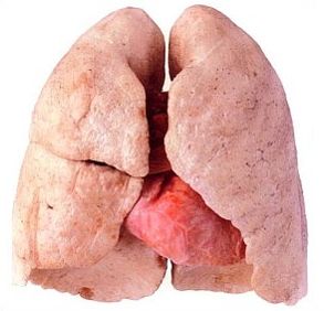
The lungs of people are the whole spineless organ. The Budova Legend was incarnated in the last century. The stench is stored from the right and left lung, rostashovuyutsya in the chest empty and to take in the main space. The most functional sign of the lungs is the participation in the gas exchange of the human body with the navkolishnim middle. The dichotomous function is to go through dichotomous paths.
Budova Legen
The skin lung is an organ that has the shape of a slightly flattened pivcone with a larger wide base (basis) and a rounded top (apex). The skin is easier to cover with its sheath - the legacy (visceral) pleura, and from the breast tissue the lungs are covered by the partial pleura (prismatic), which serves as the internal skin of the breast emptying. And in the legenevium, and in the parietal pleura, there are cells of cells, which vibrate especially in the pleural region. Tsia rіdina is found between two tsimi pleural sheaths (sheets) and "zmazu" їх, shy, dashing dick. The membranes store the pleural bear.
 The space between the sheets is called pleural empty. When the pleural emptying (pleurisy) is ignited, the pleural space is seen as a lack of quantity, it can be reduced to rubbing with leaves, and when it is dull, it appears ill-feeling... The lungs in the pleural bears are divided between themselves by the mediastinum, between them there are the heart and the great judges.
Rights і lіva legend with the same functional features are not the same for the form and size (obsyag). The middle obsyag of the stratum of people in the warehouse is close to 3 thousand cubic centimeters.
Insight into the lungs in the form and generalization of anatomical features. Pidstava (large part) lie on the diaphragm - m'yazi, like a brisket, empty from the black one, and be stored in two domes: right and left. The right dome with diaphragms is located above the pechinka, above the right part, as it is larger, and because of the whole of the left dome. Tom should lie on a new one right to the legend shorter and shorter, ale, in the middle one, 1/10 more for the general, lower left. Liva and Volodya in a lesser volume, since the heart is in the left part of the breast emptying.
Turn to list
Parts and fabrics of legends
The skin is easier to divide into parts and by segments. The right has three parts: upper, middle and lower - і ten segments. It only lasts for two parts: the upper and the lower - and is stored in nine segments. Podil on the plots of calls is designated by the spilling of large cracks: in the right door, in the left there is only one.
 Segments, how to form the legacy parts, pierced with bronchi, as they come from the new middle. It is a segmental Budova legend to build up from a great number of secondary parcels, which are stored from acinuses (in Latin “grono”). In the skin second part, there are three to five. Acinus is a structure that is even a small size, in which the process of gas exchange occurs: the blood is sour, it comes in the legend of the times, and CO2 appears, which is called when you see it. Acinusi is a functional unit of legend.
In Budov's legends, the following fabrics should be included:
- Visceral (Legendary) pleura, okremo ogorta liva and rightly easier and without getting sick, you can see pleural birth, smoothly lining of the lung with dyshly collapses along the parietal pleura in the middle of the thoracic emptying.
- Stroma (the skeleton of the lungs, which is stored from partitions, which is stored from a complete fabric). The stroma is built from fine fabrics, which are spread out on the legacy parts. In the middle of the partitions, there is the entire Legeneva "infrastructure": nerve fibers, blood vessels lymphatic systems and the roads for which to enter and go out afterwards.
- Parenchyma (tissue bean with thin skin). Legeneva parenchyma is the supremacy of all internal lung bronchial tubes and bronchioles, legeny patches, which are stored in acini, alveoli and alveolar passages.
Turn to list
Budova bronkhiv i sudin
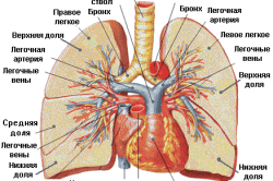 The bronchial tree is a free-flowing tube, often the ventilation system for the organisms, which repairs in the trachea, and ends in the alveoli. Visually, the bronchus buds are moving away from the tree, from the main stovbur-trachea, the head bronchi, livy and right, go out as seen in the left and right of the lungs. Then, behind the lungs, the bronchi spread out into parts, segmental, subsegmental and lobular. Large thin pieces of the bronchial tree є bronchioles, which are distributed on the endurance and endocytes of the alveolar. The structure of the bronchial tree includes alveolar passages, beetles and alveoli themselves. From the largest diameter at the point of the bifurcation (the bottom on the two nozzles) in the trachea, the distance from the ventilation tube gradually sounds, as long as it does not become microscopically thin in the alveolar passages.
The alveoli, which are located in the middle of the nighty dical canal, are of the thinnest bags in the middle, in the end they store the alveolar bears. The gas exchange is itself in the middle of the development of the legends and the gas exchange. The wall of the alveoli is a single-globular membrane shell, covered with a tissue ball, the function of which is the opening of the membrane and the appearance of the alveoli.
The membrane envelope is from the alveoli and other blood vessels - capillaries. Between the inner shells of the alveoli and capillaries in the entire five thousandth part of the millimeter. One blood-bearing capillary is suspended directly from the decilcoma by the alveoli.
 In an older person, the diameter of the alveoli becomes one quarter of a millimeter. The microscopic bags are squeezed one to one.
Capilari - the price of the least blood-bearing judges of the legends. A whole pair of organs undergoes judges of both blood circulation, small and great. In a small number of arterial arteries, they transport venous roof, and along the tributaries of veins, the arterial roof is consumed in in front of the heart s lungs. Bronchial arteries provide all necessary bronchial tubes and legenev parenchyma.
Lightly permeated with roughened hems of lymphatic vessels.
Lungs є guys organi dikhannya... The characteristic structure of the legacy tissue is laid down on another month of the intrauterine development of the fetus. Pislya folk ditiny is a diversified system of prodovzhuyu new development, the residual molding is approximately up to 22-25 years. Pislya 40-richesky vіku Leheneva fabrics for the beginning of the step-by-step start.
I will name my body in the Russian movement, having removed the authorities of power, do not drown in the water (for a rakhunok in the place of all the time). Greek word pneumon і Latin - pulmunes can also be translated as "easier". It is called “pneumonia”, which is called “pneumonia”. And for those who are ill and for those who are ill with a legacy tissue, a doctor-pulmonologist will take care of it.
Roztashuvannya
People have legends in the chest empty and borrow a large part of it. The empty chest is surrounded by ribs in front and back, the diaphragm is at the bottom. They also have a middle ground, to take revenge in the trachea, head organ blood circulation - to the heart, great (magistral) judges, stravochid and deyakis important to the structure of human body. Breast emptying does not come from the zvnіshnіm middle.
The skin of the organs is called a pleurora - a smooth serous membrane, yak has two leaves. One of them grows with the legacy fabric, the other - with the chest empty and middle. Between them, a pleural empty space is formed, filled with a small number of ridini. For the release of the negative vice in the pleural emptying and the surface tension of the line in the ny legeneva tissue, it is tightened in the straightened frame. Krim, the pleura is changing the pleura, rubbing against the rib surface during the act of action.
call of budova
Legeneva fabrics on a nagadu dibnoporous sponge of erysipelas. With wіkom, as well as with pathological processes mental systems The trivial palinny colir of the legendary parenchyma changes and grows darker.
easier maє viglyad irregular cone The top of the mountain is burnt up the mountain and is located in the area of the shoulder, standing a few centimeters across the collarbone. Below, on a cordon with a diaphragm, the Legendary surface is unmistakable. The anterior and posterior surfaces of the thigh are opaque (at the same time, the beads from the ribs are spared). The inside of the bichna (media) surface of the border from the middle and also can be seen.
On the medial surface of the cutaneous lung, there is a gate so called, through which the head bronchus and the judgment - the artery and the veins penetrate into the Legene tissue.
The two legends are not the same: right about 10% more than left... The price is tied to the rosette of the heart in the chest empty: more from the middle line of the body. The same "music" encompasses its characteristic form: right is shorter and wider, and in left - dovga and vuzka. The shape of the body lies in the form of a statue of a man. So, in artistic people, offense to the lungs of men and women, but not in healthy people, it is pummeled by the bud's breast cancer.
In the legacy tissue of people, there are no pain receptors, and the diagnosis of pain in cases of illnesses (for example, pneumonia) is linked from patients to the pathological process of pleurisy.
WHAT TO FOLD EASILY
Human lungs from anatomy are divided into three main warehouses: bronchi, bronchial tubes and acini.
Bronchi and bronchioles
The bronchi are empty tubular parts of the trachea throat and unite without the middle of the leg tissue. Head function of bronchial tubes є Air conduction.
Approximately at the level of the fifth thoracic ridge, the trachea expands into two head bronchi: right and left, which then go to the upper lungs. In the anatomy of legends more important MA system of bronchial hair removal The name of the tree is like a tree crown, so it is called “bronchial tree”.
When the head bronchus enters the Legeny tissue, it is divided into parts, and later - into more other segmental segments (according to the cutaneous Legeny segment). Further, dichotomous (guys) rose segmental bronchial tubes in the endocrine bag to bring up to the approval of bone and dichal bronchioles - found reddens of the bronchial tree.
The skin bronchus is stored in three shells:
- zovnishnyoi (from semi-fabric);
- fibrous-myazovoi (to take revenge on cartilage tissue);
- internal slime, which is covered with every part of the family.
In the world of the change in the diameter of the bronchial tubes (in the process of hair growth), the cartilage tissue and the mucous membrane gradually become aware. Found bronchi (bronchioles) do not take place at their cartilage structures, the mucous membrane is also visible. It replaces it with a thin ball of cubic drink.
acinushi
Rozpodil kintsevicheskogo bronchioles to produce until the approval of decile orders of the dichal. From the cutaneous dichal bronchioles, the alveolar walk in all straight lines, as they slowly end with alveolar bears (alveoli). The lining of the alveoli is densely covered with capillary hedgehog. It is here and gas exchange is carried out between inhaling sour and vitamins, which can be seen in carbon dioxide gas.
The diameter of the alveoli duge maliumі It grows from 150 microns in the newborn baby to 280-300 microns in the adult population.
The inner surface of the skin alveoli is covered with a special speech - surfactant. Vono pereshkojaє її decline, as well as the penetration of rіdini in the structure of the dichotomous system. Krym, surfactant can be bactericidal and take the part in the actions of the immune system.
The structure, up to the warehouse, which includes the dichal bronchiole and the alveolar walk and the beetles, is called the primary part of the leg. It has been established that approximately 14-16 chillers can enter from one of the bronchial tubes. Otzhe, such a number of primary parts of the lung fix the head structural unit of the parenchyma of the lung tissue - acinus.
I named it anatomical and functional structure through a characteristic znichnogo viglyada, scho grono grapes (Latin Acinus - "grono"). In the body of people there are about 30 thousand acini.
The back of the area of the dikhal surface of the leg tissue behind the alveolar opening ranges from 30 sq. meters at vidihu and up to approx. 100 sq. meter with inhalation.
SHARES I LUNG SEGMENTS
Acinushi to mold particles Who pretend to be segments, A z segments - parcels, Warehouses are easier.
In the right legacy there are three parts, in the left one - two (through a smaller size). In both legends, the upper and lower parts are seen, and on the right, the middle and middle parts. Between themselves the parts were made by furrows (fisurs).
parcels addicted to segments, I do not conceal the visible space between the eyes of the semi-woven fabrics. chime in the right legend there are ten segments, in the left - visim... The leather segment should be placed in its warehouse for segmental bronchus and a type of leg artery. Zovnishny viglyad The Legendary segment has a similarity with the wrong form, the top of which is turned to Legendary collars, and at the beginning - to the pleural leaves.
The upper part of the cutaneous lung is the anterior segment. In the right leg, there is also an upper and a posterior segment, and in the left - an upper-posterior segment and two lingual (upper and lower).
In the lower part of the cutaneous lung, the upper, anterior, lateral and posterior basal segments are developed. Krym tsyogo, in the legends of the beginning of the mediobasal segment.
The middle part of the right legacy has two segments: medial and lateral.
For segments of the lungs of people, it is necessary for a clear localization pathological changes Legendary textiles, which are especially important for practicing drugs, for example, in the process of getting rid of and guarding against pneumonia.
FUNCTIONAL RECOGNITION
The main function of the legends is gas exchange, when one can see carbon dioxide gas from the blood in one hour sour, necessary for normal metabolism of practically all organs and tissues of the human body.
When we breathe in, we sour Witnessing along the bronchial tree penetrates into the alveoli. There you will come "vidpratsovana" shelter from a small amount of blood circulation, as to take revenge on a large amount of carbon dioxide. By sending the gas exchange to the carbon dioxide, the gas is known again along the bronchial tree to be named when you see it. And we have a sour shelter to come to a great blood circulation and go far to the organs and systems of the human body.
The act of dikhannya among people є mimovilny, reflexive... The structure of the brain is special for the process. prodovuvaty brain(Dichny center). Behind the step of saturation of blood in carbonic acid, the rate and glybin of energy are regulated, as it grows more and more often when the concentration of gas is increased.
The lungs have no meat tissue... To this, his fate in the act is viciously passive: expanding and squeezing when the breast tissue collapses.
Have zd_ysnenna dikhannya take the fate m'yazova fabric diaphragms and breast cells. Apparently, I see two types of behavior: the chest and the chest.
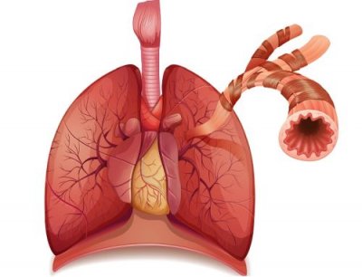
On the inhale, the volume of the empty breast is growing, in niy a negative grip collapses(Lower than atmospheric), as you allow me to go over and over again in the legend. Do not hesitate to use fast diaphragms and m'yaz frame breast cells (mid-ribbed muscles), which should be produced until the ribs are raised and loose.
On vidikha, navpaki, the grip of the atmosphere, and the vivedennya of the food saturated with carbonic acid is carried out practically in a passive way. With a large amount of emptying of the chest, it will change for the opening of the weakening of the spontaneous muscles and the lowering of the ribs.
With some pathological camps, the act of dichannia includes and so-called additional dichalny m'yazi: shia, chernogo press, etc.
Some of it, like a person inhales and vidikhaє at a time (dichal volume) to become close to a pivot. In the middle of the day, a chilin is seen in the middle of 16-18 dykhalnye rukhiv. For dob, pass more through the Legeneva fabric 13 thousand years!
The average mix of legends becomes approximately 3-6 liters. At the people of Nadlyshkov von: for an hour I vikorovuєmo just close to one eighth part of the community.
Krim gas exchange, in the legends of people є y іnshі functions:
- Participation in the acid-puddle balance.
- Vivedennya toxins, essential oils, Steam alcohol і t. D.
- pidtrimka water balance organism. At the norm, through the legends of viparovutsya close to a glass of water for good. In extreme situations, additional water supply can be up to 8-10 years.
- The state of the art of clearing and detecting conglomerates of clitin, microembol fats and fibrin clots.
- Participation in the processes of blood circulation (coagulation).
- Phagocytic activity - participation in the robotic immune system.
Otzhe, budova and funktsii it is the legend of people to be in close interconnection, so that they allow the robot to be insensitive to all human organisms.
Know the pardon? View and Print Ctrl + Enter
Easy people є one of the most important organs, without any ill-feeling. Dikhannya is created for us so natural, but for the most part, in our body we see folding processes to make our life unchanged. To be more intelligent, it is necessary for the nobility of the Budov legends.
In the process, the energy goes through two bronchi, which may Riznu Budova... Livy is better than the right, a little more, the more often alien tilo penetrates through the right bronchus. Tsі organi cant distribution. When you enter in the easy right turn into 3, and livy into 2 parts, so you can see a number of parts of the legends.
Budova legends to reach the fold, the fragments of the bronchi in the middle of them are distributed on the empty other segmental bronchi. At its own decks, they pass into the lobular bronchi, scho enter the legs to a small part. It’s important to see, like Budova legends, I don’t know, there are some small bronchial tubes in them (there are close to 1000). The intra-lobe of the bronchi can be up to 18 hiloks (thermal bronchioles), which do not grow in their cartilages. Tsіntsevі bronchіoli establish the structural storage of the lungs - acinus.
Budova's legend is simpler to learn, sounding, which itself is acinus. Qia is a structural unit of suction of alveoli (lost respiratory bronchioles). Їх walls are a material substrate for gas exchange, and the area for an hour of repeated inhalation can be up to 100 sq. M. The most stretching of the psychic surfaces is displayed during the physical days.
The bronchopulmonary segment is called a part of the legacy part, it is ventilated by the bronchi of the 3rd order, it is seen from the bronchus. Skin from them I will okremu broncho-sudinny nіzhku (artery and bronchus). Segmental Budova legend has been revealed in the course of development of medicine and surgery. In the right legacy there are 10 segments, and in the left one - 8. Because of the establishment of pulmonary growths on the bronchopulmonary segments, it became possible to see the growth of the lungs in the lungs with the maximum protection of healthy parts. 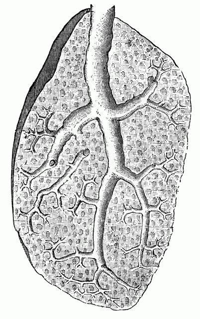
At the same organ, it is accepted to develop such surfaces: middle, diaphragmatic, rib. In the middle, this is the name of the “gate”. Through them, the bronchi, arteries and nerves enter the legends, and the lymphatic judges and the legene veins enter the legends. All tsi affirmed to become so the title of "root of the lung."
Easy to grow by the grooves of the growth and triviality. The stench podilyayut fabrics to the very doors of the legends. Razrіznyayut 3 parts of the right legion (lower, upper, middle) and 2 left (lower, upper). The lower parts are the largest.
Budova legend will be incomprehensible without urahuvannya visceral leaves of the pleurisy, which cover the skin legend and the region of the root and make a "partial leaf", which whistles the walls of the breast emptying. Between them, a slit-like empty space is roasted, a part of which is called sinuses (rostered between the parietal leaves). The most costal-diaphragmatic sinus is inserted into the most pleural sinus (the edge of the lung descends into the new one when inhaling).
Budova Legen will explain the processes, which are seen in them before the hour of the dichotomy. In the whole body there are 2 systems of blood-bearing vessels: small (to be stored from the veins and arteries, to take part in gas exchange), great (to be stored from bronchial arteries and veins, but to deliver arterial blood for the loss of pain Behind the character of their habits, they are similar to the artery, but they do not see their own nonpotency. Їх dzherelom є capillary hedgehogs, interlobular tissues, other bronchial tubes and visceral pleurisy. Interlobular veins are formed from capillary hemlines, which are angry with themselves. Behind them there are more veins that pass near the bronchial tubes. Two veins and segmental veins in the cutaneous lung are formed by two veins: lower and upper (their size is very variable). The smell is okremo fall into the front of the heart. 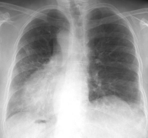
The number of bronchial arteries is not permanent. It counts from 2 to 6. In 50% of vypadki in humans there are 4 bronchial arteries, which go equally to the left and right head bronchi. The stench is not є in the arteries of the bronchial tubes, the splinters go out to the middle organs. The ear of the right arteries grows in the cells behind the trachea and in front of the trachea (between the lymph nodes). There are arteries of rosette in the cells of the lower trachea and under the arcuate aorta. In the middle of the lung artery is located in the cells of the bronchial tubes and, in the middle of the lungs, playing a role in the blood loss of the first parts of the pleurisy. In the respiratory bronchioles, the stench absorbs its own meaning and passes into the capillary system.
All lungs are tied one by one. The extra-organ and internal anastomoses are created to create an insult to the blood circulation.
The lymphatic system consists of cob capillary heels, gossiping in the middle of the body, in the outer vessels, in the lateral and in the inner lungs in the lymph nodes. They develop superficial and glibody lymph judges.
Dzherelo іnnervatsії legends - nerves gossip and storms of the middle, approved with gilkami cute, bluish, spinal and diaphragmatic nerves.
Lungs - tse the organization of dichannya, in which gas exchange is carried out between the witnesses and the circulatory system living organisms. Lungs in savts (before them, ludin), plasuns, birds, in most species of amphibians and some species of ribs.
The name of the tsikh organs is unavoidable. If people crumbled the carcasses of food and put the entrails from them into a basin of water, then all the organisms appeared important for the water and sank to the bottom. Only the organizing action, roasting in the breast, the bullets for the water and swam on the surface. So the name "lungs" was attached to them.
And for that, as they briefly sang, it is also easy, let us wonder, it is also the legends of people and the stench of vashtovani.
Budova legeniv people
Lungs are a great organ. The skin man has two lungs - right and left. Easy rosetting in the chest and occupy 4/5 of the year. The skin is lightly covered with pleurora, the lasting edge of which is breast tissue... A spate (in new-born women) is lightly bloody-rhymed colir. From the plinth of life, light step by step darken through the accumulation of particles of wood and saw in them.
Skin is easier to fold in a part, rightfully, there are three parts, and it’s just two parts. Parts of the lung are divided into segments (10 in the right legend, 8 in the left), the segments are folded into small parts (there are nearly 80 pieces in the skin segment), and the small parts are added to acini.
Drinking in the legends through the wild throat (trachea). The trachea is divided into two bronchi, the skin is included in the legend. Distant skin bronchus extend according to a tree-like principle to the bronchi of a larger diameter, which leads to the dermal part, skin segment, skin part of the leg. The bronchus, which is included in the part, lasts for 18 - 20 bronchioles, the skin of which ends with ACINUS.
In the middle of the acinus of the bronchial tubes, they spread on the alveolar path, all the alveoli. The alveoli are wrapped around by a net of thin blood-bearing vessels - capillaries, from the alveoli of the naythonshoy stinka. The very center of the alveoli is supplied with gas exchange between the blood and the witnesses.
Yak start legends
On inhalation from the trachea through the fringe of the bronchial tubes and the bronchial tubes are consumed in the alveoli. From the іnshy side to the alveoli along the capillaries to come up with a roof, overpopulated with carbon dioxide gas. Here the shelter of the people is cleaned up in carbon dioxide and sourced for the needs of the client's body. In the presence of carbon dioxide, the gas from the lungs wicks into the atmosphere. The whole cycle repeats itself indefinitely, until the end of life.


