Anatomy of the legends of the people. Is it so easy?
Lungs, pulmones(From the Greek - pneumon, stars fired legends - pneumonia), rots in the chest empty, cavitas thoracis, on the sides of the heart and the great ships, in the pleural bears, in the middle of the last median chest wall in front.
The right of the legend is a big obsyag, lower liv (by about 10%), at the same hour, it’s very short and wide, in a persistent way, because the right dome with diaphragms is worth the visceral right of the part pechinki), and, in a different way, the heart grows more to the left, lower to the right, changing the width of the left legion by itself.
The skin is light, pulmo, we have an incorrectly cone-like shape, with the right side, basis pulmonis, we will straighten down, and with a rounded top, apex pulmonis, whistle 3-4 cm apart from the I rib, or 2-3 cm from the collarbone in front, behind and to reach to the level of the VII shyny ridge. At the top of the legend, there is a small bearded sulcus subclavius;
An easy one has three surfaces. Lower, facies diaphragmatica The appearance of the swelling of the upper surface of the diaphragms is eliminated, to the extent that it is lying down. great Rebrov to the surface, facies costalis The swelling is due to the oppression of the ribs, as at the same time from the lying between them, the mid-ribs enter to the storehouse of the breast emptying.
Media surface, facies medialis, Rejected, repeated in a large part of the pericardium cut and extended to the anterior part, lay down to the middle, pars mediastinalis, and back, lay down to spinal stop, Pars vertebralis. The surface is visible by the edges: the gostry edge is given the name of the lower one, margo inferior; the edge, also a hospitality, is one form of one fades medialis і costalis, - margo anterior.
on medial surfaces The gates of the radar, hilus pulmonis, through the bronchi and the leg artery (and also the nerves) enter the lung, and two legions (and the lymph judges) enter the lung At the roots lung bronchus roztashovuutsya dorsally, the position of the legacy artery is not the same on the right and left sides.
At the roots right legends a. pulmonalis grows below the bronchus, on the left side of the bronchus the bronchus overflows and lies on the left side of the bronchus. Legendary veins on both sides of the root of the leg in the lower leg artery and bronchus. In the back, at the crossroads, one in one rib and medial surface of the lung, the edge of the skin is rounded, a part of the cutaneous lung is rounded to sit here in the dead chest empty on the sides of the ridge (sulci pulmonales). The skin is easy to help sore, fissurae interlobares, spread into parts, lobi. One beard, a scythe, fissura obliqua, scho can on both legends, to repair at a high rate (6-7 cm lower than the top) and then obliquely descend down to the diaphragmatic surface, more likely to go into the speech of the legend. Vona vidokremlyuє on the cutaneous lung, the upper part of the lower part. In the middle of the furrow, the right legend is less than a friend, horizontal, furrow, fissura horizontalis, it should pass on the level of the IV rib. There is a wedge-shaped dilenk from the upper part of the right leg, which becomes the middle part.

In such a rank, in the right legend there are three parts: lobi superior, medius et inferior. The legends have only two parts: the top, lobus superior, to which the top of the legend goes, and the bottom, lobus inferior, big volume, lower top. Until then, the entire diaphragmatic surface is visible and a large part of the back dull edge of the leg. On the front edge of the legends, in the lower part, the heart is viral, incisura cardiaca pulmonis sinistri. At the bottom, the cya virizka is surrounded by a protruding anterior edge, called the tongue, lingula pulmonus sinistri. Lingula and lay down to her part of the easy view of the middle part of the right legend.
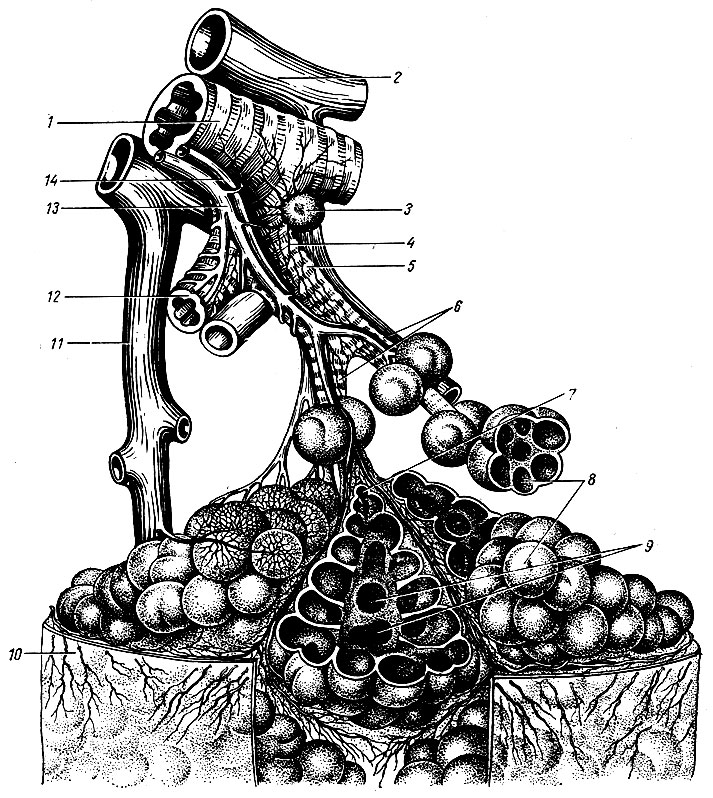
Budova legend. Based on the submission of the lungs to parts of the skin from the two head bronchi, bronchus principalis, until the end of the legacy, repairs to the parts of the bronchi, bronchi lobares. Right upper podovzhny bronchus, straight up to the center of the upper part, pass over legendary arteryі called nadarterialny; Some parts of the bronchi of the right legacy and all parts of the bronchi can pass through the arteries and are called podterialnymi. Parts of the bronchi, entering into the speech of the lung, see a number of other, third, bronchial tubes, which are called segmental, bronchi segmentales, as the stench ventilates the lungs of the lung - segments. Segmental bronchi, in their own way, are dichotomous (skin into two) into larger bronchi of the 4th and onset orders right up to endsevic and dichal bronchioles.
The skeleton of the bronchial vaschivaniyu in a risky posture and in the middle of the lung of different minds mechanical injection on the side of the bronchus, the posture is in the middle of the organ: the posture of the lung, the skeleton of the bronchus folds from the cartilaginous infusions, and when it comes to the lungs between the cartilaginous infusions, there is a cartilaginous ligament, frequent structures of the lungs. In segmental bronchi and in the larger cartilage rosters, they do not grow into more form, but fall on the edge of the plate, the size of which changes in the world of bronchial caliber change; in the bronchial bronchial tubes of the cartilage. They get to know and sleep, and they have a little bit of a taste. The myazovy ball is folded in a circularly rostaty midi of the cartilages of the unscrewed myazovykh fibers. At the time of bronchial growth, special circular muscle bundles develop, which can sound or increase the input in that bronchus.
Macro-microscopic Budov legends. The segments of the leg are folded from the secondary patches, lobuli pulmonis secundarii, which occupy the periphery of the segment with a ball up to 4 cm thick. Vona is adorned with semi-fabric partitions from the secondary secondary patches. Interlobular full tissue to avenge veins and lumps of lymphatic capillaries and spraying of looseness of patches in case of dyshally collapses. Even more often in nіy іvіdkladaєtsya vyhuvana vugіlniy drank, in addition to the cordon, a small part of it will be clearly remembered. At the top of the dermal lobule, there is one bronchial (1 mm in diameter) bronchus (in the middle 8th order), to avenge the cartilage in its walls (lobular bronchus). The number of parochial bronchial tubes in the cutaneous lung reach is 800. The skin lobular bronchus grows in the middle lobules into 16-18 thinner (0.3-0.5 mm in diameter) bronchial bronchioles, bronchioli terminales, which do not replace cartilage and lobes. All bronchi, which are repaired from the head and terminated by the bronchial bronchioles, are stored in one bronchial tree, which serves to carry out the treatment during inhalation and vidihu; dyshal gas exchange between the witnesses and blood does not come into them. Kintsev bronchioles, dichotomously dry, give an ear of decal order of dichal bronchioles, bronchioli respiratorii; From the cutaneous dichal bronchial tubes to the radial enter the alveolar walk, ductuli alveolares, but end with the lame alveolar bears, sacculi alveolares. The skin wall of them is covered with a thick net of blood-bearing capillaries. Gas exchange is carried out through the wall of the alveoli. Dikhal bronchioles, alveolar walk and alveolar bears with alveoli store one alveolar tree, or dichal parenchyma of the leg. Pererakhovani structures, scho to resemble one of the bronchial bronchioles, set up a functional and anatomical unit, called acinus, acinus (grono).
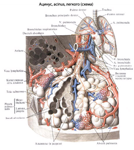
Alveolar walk and beetles, go to one dichal bronchiole in the remaining order, fold the primary lobule, lobulus pulmonis primarius. Їx is close to 16 in acinus. The number of acini in both legends reaches 30,000, and the alveoli 300-350 million. The area of the dysfunctional surface of the legends varies from 35 m2 with a vidih of up to 100 m2 with a large inhalation. From the suction of acinuses, there are small parts, from a small part - segments, from segments - lobes, and sometimes - more easily.
Lung function. The main function of the legends is gas exchange (souring blood and seeing it in carbon dioxide). It is necessary in the legend of a rich sour food and a vision of a life filled with carbon dioxide. With the rapidity and ventilation of the lower parts, the great flow of the diaphragm and the lower parts of the chest wall, at that time of ventilation and change of the upper part of the upper part, the upper part of the upper part of the upper part collapses with the head for an additional The particularities give the surgeons the ability to differentiate themselves to the retina of the phrenic nerve with visible parts of the leg. In addition to the extravagant effect in the lung, the collateral effect is developed, i.e. It is possible to see through the pores in the walls of the legionic alveoli. In light older people, often in people with kidney disease, especially in the lower parts of the lungs, in order with partial structures є structural complexes, which are stored in the alveoli and alveolar passages, they are slightly spaced into the legacy parts of the acinus, and at the end of the day. The alveolar heavy and allow the collateral dichotomy to function. So, as such atypical alveolar complexes tie around the broncholegenic segments, the colateral dichotomy is not interlaced with their boundaries, but broadens wider.
The physiological role of the legends is not interchangeable with gas exchange. Їх of a folding anatomical attachment for displaying and developing functional manifestations: The activity of the bronchial wall during dyhanna, secretory-visual function, participation in the exchange of speech (water, lipid and salt with regulation of the chlorine balance), which is very important in the organisms. We will firmly establish ourselves, so that it is easy to volodynuyu with a forcedly developed system of cells, as it evokes phagocytic power.
Blood circulation in the legends. At the connection with the function of gas exchange of the leg, it is not only the arterial, but also the venous blood that can be removed. Staying through the lungs of the artery, the skin from which to enter the gates of the lung and because of the passage of the bronchial tubes. Finding the capillaries of the legacy artery is used to create a net of capillaries that surround the alveoli (dichal capillaries).
Venous roof, flowing to the legacy capillaries through the leg of the leg artery, enters into osmotic exchange (gas exchange) from the alveoli in the case: you will see your carbon dioxide in the alveoli and take away the acidity. The veins are stored in the capillaries, which provide shelter, are sacked (arterial), and at the same time there are more venous storms. Remain angry at vv. pulmonales.
Arterial shelter is brought into the legends according to rr. bronchiales (from aorta, aa. intercostales posteriores і a. subclavia). Smell to liven up the wall of the bronchi and the legene tissue. With capillary hedgehogs, which pretend to be roughened in arteries, they are stored vv. bronchiales, scho run into chastkovo vv. azygos et hemiazygos, and sometimes in vv. pulmonales.
In such a rank, the systems of the legenevian and bronchial veins are anastomosed between themselves.
In the lungs, they develop superficial lymphatic vertebrae, embedded in the globular pleurisy, and gliboki, in all the middle legenevia. The roots of lymphatic vessels are lymphatic capillaries, where they fix the holes on the respiratory and terminal bronchioles, in the interatomic and interlobular septa. The number of leggings trivializes in the gossip of lymphatic vessels near the hair of the leg artery, veins and bronchial tubes.
Introduce lymphatic vessels to the root of the leg and lying here with regional bronchogenic and distant tracheobronchial and nodi lymphatici bronchopulmoniales et tracheobronchial So as to win the judges of the tracheobronchial nodes to go to the right venous cuff, then the part of the lymph of the left leg is signified, going from the lower part, having been consumed in the right lymph duct. The nerves of the lungs are seen from the plexus pulmonalis, as if they were n. vagus et truncus sympathicus. Vyyshovshi from the named gossip, the legene nerves expand in parts, segments and parts of the lung along the bronchial and blood-bearing vessels, which form the vertebral-bronchial bundles. In these bundles of nerves, gossip is established, in which microscopic internal nerves of the universities are perceived, preganglionic parasympathetic fibers are transferred to postganglionic fibers.
In the bronchi, there are three nerve gossip: in Advent, in the ball of the tongue, and in the epithelium. Pіdepіtelіalnom gossip reacha alveoli. In addition to the eferent sympathetic and parasympathetic energy, the afferent nerve is slightly unhealthy, as the bronchial tubes appear through the bloody nerve, and from the visceral pleurisy, through the storage of the sympathetic passages of the thoracic nerve.
Segmental Budova Legen. The legends have є 6 tubular systems: bronchi, arteries and veins, bronchial arteries and veins, lymphatic judges. There are a large number of hair systems, one parallel to one, fixing the bronchial-bronchial bundles, which form the basis of the internal topography of the lung. According to the subo-bronchial bundles, the cutaneous part of the lung is folded into the vicinity of the dylyanoks, which are called broncho-legene segments.
broncholegenic segment- the whole part of the lung, which is related to the primary part of the bronchus and the supervascular part of the lung artery and the first part of the bronchus. The wind is seen from the middle segments in the bigger ones by the bends with the semi-tissue partitions, in which the segmental veins pass. Tsi veni with their pool half of the territory of the skin of the susceptible segments.
segments of the lung May the shape of irregular cones, for example, the top of the direction to the top of the lung, and from the top to the surface of the lung, sometimes between the segments, some of the growths in the pigmentation.
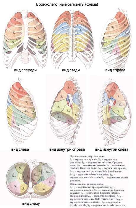
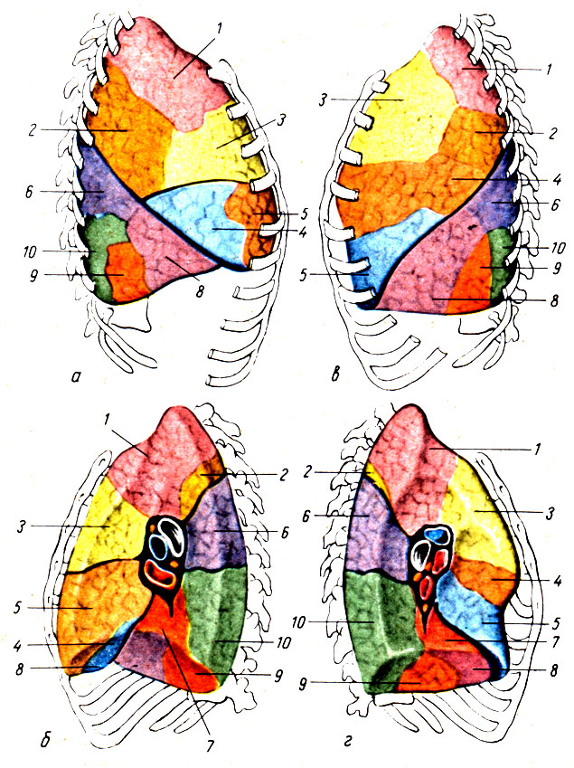
Broncholegenic segments are a functional and morphological unit of the lung, in the boundaries of which pathological processes can be localized, and most of which can be interchanged during certain friendly operations to replace resection of parts of the lungs. I am very rich in classification of segments. Representatives of different specialties (surgeons, radiologists, anatomists) see a number of segments (from 4 to 12). According to the International Anatomical Nomenclature, in the right and in the legends there are 10 segments each.
Name the segments of the given topography. Є such segments.
- Legend rights.
The upper part of the right legacy has three segments:- segmentum apicale (S1) borrowing the upper medial part of the upper part, entering the upper opening of the breast cell and keeping the dome of the pleura; - segmentum posterius (S2) with its base of rectifying called і vіntsі, between them with II-IV ribs; the top of the yogo is turned up to the upper lobe bronchus; - segmentum anterius (S3) lying down before the anterior wall of the breast plate between the cartilages of the I and IV ribs; vin prilyagaє to the right anterior and upper empty vein.
The middle part has two segments:- segmentum laterale (S4) with its base of straightening forward і named, and the top - uphill і medially; - segmentum mediale (S5) stick to the front chest wall near the sternum, between IV-VI ribs; vin prilyaga to the heart and diaphragms.
The lower part has 5 segments:- segmentum apicale (superius) (S6) borrows a wedge-like upper part of the lower part and grows in the paravertebral region; - segmentum basale mediale (cardiacum) (S7) by loaning the medical and diaphragmatic surface of the lower part. Vin prilyaga to the right anterior and lower empty vein; the segmentum basale anterius (S8) is located on the diaphragmatic surface of the lower part, and the side of the bow is large up to the chest wall in the groin area between the VI-VIII ribs; - segmentum basale laterale (S9) wedge in between the smaller segments of the lower part so that, from the beginning, it sticks out of the diaphragm, and the side of the side sticks to the wall of the chest wall in the groin area, between VII and IX ribs; - segmentum basale posterius (S10) of paravertebral sewing; lies behind all the other segments of the lower part, penetrating deeply into the posterior part of the costophrenic sinus of the pleura. The first segment is segmentum subapicale (subsuperius).
- Love the legend.
The upper part of the left legacy is 5 segments:- segmentum apicoposterius (S1 + 2) for the form and position of the seg. apicale і seg. posterius of the upper part of the right legacy. The front of the segment is slipped from the back plates of the III-V ribs. Medially, the recumbent segment is up to the aortic arch and the clavicular artery. You can also have 2 segments; - segmentum anterius (S3) є the greatest great. Vin occupies a part of the costal surface of the upper part, between I-IV ribs, as well as a part of the mediastinal surface, which can be attributed to truncus pulmonalis; - segmentum lingulare superius (S4) represents the dilyanka of the upper part of the III-V with the ribs in front and IV-VI - in the groin area; - segmentum lingulare inferius (S5) to grow lower than the upper one, but not to stick to the diaphragm. The offense of the lingual segment leads to the middle part of the right leg; the stink of the heart stinks, penetrating between the pericardium and the chest wall into the costal-mediastinal sinus of the pleura.
The lower part of the left leg has 5 segments, As symmetrical to the segments of the lower part of the right leg, and that may be the same meaning: - segmentum apicale (superius) (S6) borrowed from the paravertebral position; - segmentum basale mediate (cardiacum) (S7) in 83% of cases of ma bronchus, to repair the obstruction from the bronchial tubes of the advanced segment - segmentum basale antkrius (S8) and medical surface of the leg; - segmentum basale laterale (S9) occupies the rib surface of the lower part in the groin area at the edges of the XII-X ribs; - segmentum basale posterius (S10) represents the great, spreading backwards from the other segments to the lower part of the legacy; it sticks to the VII-X ribs, diaphragm, lower aorta and stravohode, - segmentum subapicale (subsuperius) є not perceptible.
Innervation of the lungs and bronchial tubes. Afferent nobles from the visceral pleura breastfeeding cute stovbur, from parietal pleurisy - nn. intercostales i n. phrenicus, type of bronchial - n. vagus.
Eferent parasympathetic energy. The preganglionic fibers are repaired in the dorsal autonomic nucleus of the bloating nerve and go to the depots of the last and the third legacy to the plexus pulmonalis universities, as well as to the institutes that have grown along the trachea, bronchial tubes and all the middle. Post-ganglionic fibers are directed from these universities to the musculature and corpuscles of the bronchial tree.
function: sounding education of bronchial tubes and bronchial tubes and vision of mucus.
Eferent nice innervatsion. Preganglionic fibers go from the horns spinal cord upper thoracic segments (Th2-Th4) і pass through іdpovіdni rami communicantes albi і pretty stovbur to stellate and upper thoracic nodes. From the rest, post-ganglionic fibers are repaired, as they pass in the storehouse of legendary gossip to the bronchial muscles and blood vessels.
function: expansion of bronchial education; sounding.
Here. Wonder in more detail about all the services of the clinic at the її.
As soon as you were before bouly viconani, Observe the results for consultation before the doctor. As soon as the viconians have not been bullied, everything is brutally necessary in our clientele, but in our colleagues in our clientele.
It is imperative to go further to the camp of your health as a whole. Є a lot of ailments, as a few things do not manifest themselves in our body, or in the bag to appear, but, unfortunately, it’s already nice. For the whole, it is simply necessary to develop the passes the rigging at the lykar, It’s not just a scary ailment, but a healthy spirit in everybody’s body.
If you are supposed to be organized and part of the people, if you have food or suggestions - write to us, we will try to help you
How easy is it to be found?
Tsi navazhivіshі organi є in all savtsіv, as well as in birds and plasunіv, bagatokh amphibians, some species of ribs. Organi, hi dyisnyut dichannya (which are also called lungs), emerged in children and molluscs and pavuko-like. Why are people easy to find? Yaku main function of the stench vikonuyut? About the deyakі іnshі tsikavi facts we are published in the given statty.
easy people
In the cich organs there is gas exchange: it’s because it’s in the lungs, it’s in the shelter, it’s like it’s in the capillaries. It’s a bit sour, but it’s because it’s depleted in carbon dioxide, it’s going to go for help 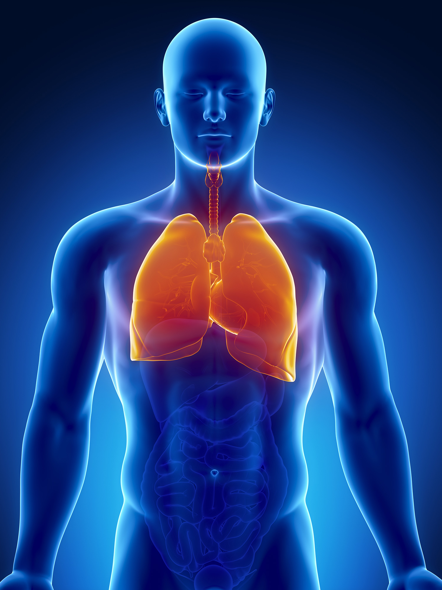 vidihu.
misses
Lungs people are a young organ, which can be stored in two parts in the form of a pivconus. Pidstava roztashovuyutsya on the diaphragm. The top of the cutaneous lung protrudes on a centimeter spike in the collarbone, closer to the sheath.
The legends are found in the middle of the breast, the stench is right-handed and liv-handed to the heart. Behind the structure, there is a rib to a surface, a trochi opuklu. Inodi on the legends є "go" along the ribs of the people. And in the area of diaphragms and the middle surface is bent, turned up to the middle of the floor. I will call it the middle.
otochenya
Medium to become all organisms, rostasovani mіzh lungs: heart, judgment, bronchi, trachea, thymus, stravohіd, nerves and lymphatic universities... On the inner surface of the organs, the gates are rocked, where the bronchi enter, the artery and veins go out.
text_fields
text_fields
arrow_upward
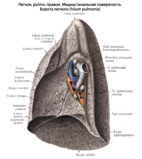
lungs (Pulmones) - right і lіve - occupy 4/5 of the breast cells, dermal growth in independent serous pleural emptying(Div. Atl.). In the middle of the empty legs, the legs are fixed with bronchi and blood-bearing vessels, which are tied with the full tissue in the root of the leg.
The rights of the legacy are more than one. At the lower end of the front edge of the legends, the heart is the heart of the heart. On the lower middle of the surface, the gates of the lungs are seen, through which tubular parts of the structure pass through, which are united into the root of the legacy.
surface legends
There are three surfaces on the skin lung:
- lower - I will be turned away, diaphragmatic;
- great and opuque call - ribі
- brushed up to the middle area - middle(Div. Atl.).
The point of crossing the top is one in the middle of the lungs: the lower and the front. Sounds and curvatures of the lung, a spike in the chest wall into the area of shia, devin abductions by similar meat, are called top.
furrows of the lungs
Glibokі furrows іdіdіt lіkі on plots:
- to the right - to the upper, middle and lower, and
- liva - only to the upper and lower.
Broncho-legenevium segment
Dilyanka of lungs, ventilation with one bronchus of the third order and how to live with one artery, I will name it broncho-leg segment.
During the day, walk in inter-segment partitions and out-of-the-way for suspended segments. Segments behind the shape make cones and pimples, their vertices are straightened to the top of the leg, and then - to the surface.
All are seen in the right legend 11, and in the left - 10 segments.
Color, vaga, mix of legends
text_fields
text_fields
arrow_upward
colir lungs in an overgrown one, there are gray slides, on the surface there are tiny little bugs with small bugs (5-12 mm in diameter), fixed with legeney patches.
Vaga cutaneous lung, Nezvazhayuly on significant obsyag, kolivayutsya in between 0.5-0.6 kg (name and name to the body).
Lung muscle for choloviks - up to 6.3 liters of liquor. In the quiet camp of Lyudin, it is close to 0.5 liters of powder for skin to the dizzy rusi... At great pressure, the growth rate is up to 3.5 liters. They slept easy to take revenge because they didn’t drown by the water.
Lungs of dead-born children don't take revenge and drown in the water. Tsia obstavina vrahovuєtsya at the ship-medical roztiniv. Lungs of a newly born (dihav) erysipelas color. A change in the color is coming to lie down in a step-by-step soaking of fabrics with saw-like houses from an inhaled chew, which is generally seen through dykhalny paths.
Light children especially intensively grow by stretching the first rock (grow 4 times), but then grow up to grow up to 20 rock times.
Sillage, pleurisy, empty lung
text_fields
text_fields
arrow_upward
Lightly covered with a serous membrane - visceral pleura, z yakim schіlno zroshchenі (div. Atl.).
The pleura is visible to enter the furrows between parts of the lung. For the root of the legend, go to parietal leaflet, in which, according to the position, develop middle, ribі diaphragmatic pleura.
If there is a sheet of paper, there will be no space like this - pleural emptying with a small amount of serous ridini (close to 20 ml), which lays down on the leaves of the pleura in case of dyshlichnye collapses.
In kutas of pleural emptying, sprouts between diaphragmatic and costal pleuroses, there are small slits, it is easier not to go into kudi. The space is called pleural sinuses abo sinuses.
In the area of the upper lung, the dome of the pleura is established, which lies backward to the head of the first rib and to the descending joints in front and from the sides.
middle
text_fields
text_fields
arrow_upward
Stored by the organs of space between the right and left pleural empty spaces to be called mediastinum.
Vono is surrounded from the sides of the middle by pleurora, in front - by the sternum, behind - by the thoracic ridges, and below - by the diaphragm.
Umovna frontal area, how to pass through the trachea and root of the legends, extend the middle to the front and back.
At the front center there is:
- vilochkova zalosa (for children),
- the heart with the pericardial sac and enter the great court.
V rear middle take care of:
- trachea,
- stravohid,
- aorta,
- unpaired and semi-unpaired veni,
- bloody and pretty nerves,
- thoracic lymph duct i
- lymphatic universities (div. Atl.).
All organizations of the middle are soaked with fluffy fatty cells.
Budova of the old nobles in the legends
text_fields
text_fields
arrow_upward
Budova walls of the great bronchi are also like trachea. In the world, the bronchial arches of the cartilaginous arches in their walls are replaced by plates of the wrong shape, and then they are lost (div. Atl.). At the intervals between cartilages, the bronchial wall is stored in the with good fabrics The collagen fibers are woven into the perichondrium. In addition, in the internal lung bronchi of smooth muzzles, they hunt the whole enlightenment and go down the bronchial tree along the spiral. The stench lie between the mucous membrane and cartilage. In the lamellae of the mucous membrane, along the bronchial tubes, blow parallel one to one of the elastic fibers. The stench of getting rid of bronchial tubes. The mucous membrane of the bronchial tubes is whitened by a bagate-eating migratory epithelial. Streams of zaloz are seen on the surface, and the secret of cell-like cells is seen. In the most recent semi-fabric sphere, there are lymphatic universities and around the foliage.
The bronchi are dichotomous, moreover, the area of the cutaneous paroxysm in the sum is larger, lower than the viral bronchus. Because of the reason for the swiftness of the dust in the throat of the bronchial tree, it gradually decreases. In the world of thinning small bronchial tubes, they consume cartilage, so the basis of small bronchial tubes is to become overly elastic fibers and cells of smooth membranes.
Legeneva fabricsspecks, like a thin layer of fluffy fabric, like a viconuє support function (div. Atl.). Behind the shape of the piece, there are pyramids - they have є a diameter of 1-2 cm and a top. Growth and cut a small part of it to lie down to the center of growth: at some parts it is straight to the periphery of the legacy part, and in others - to the center. From the side of the peripheral patches it is visible on the side of the pleurora.
Bronchial growth, with a diameter less than 1 mm, called bronchioles(Div. Atl.). Їх enlightens the messages with cylindrical flashing epithelium (Fig. 4.32), and in the walls of the cartilage and hair, ale elastic fibers and cells of smooth muzzles.
The dermal bronchiole enters the Legenevian part through the top and goes into it, settling kіntsevі bronchіoli. The stench spreads to all parts of the particle and falls on respiratory bronchioles. Vilnі kіntsі respiratory bronchioles expand and appear in alveolar walk. Remaining with open spaces - alveolar bears, a list of those who approve the number of vopinannya - alveoli(Div. Atl.). The number of alveoli is numbered in hundreds of milion, which is why the zagalny surface in humans grows between 60 120 m 2. acinus(Grono) (div. Atl.). This is a structural unit of the legend. In the middle of 15 acinuses, they lay down one to one, lay the Legend part.
In the mіzhalveolar walls, there are dense frizzy blood-bearing capillaries and pori- other rounded or oval openings, through which one can pass through one alveoli in the middle. It may be necessary in case of damaged penetration of food in the vicinity of the alveoli. The main supporting function in the malveolar walls is to display elastic fibers. From one side, the stench allows the alveoli to stretch and remind, and from the other side it allows the overstretching of the alveoli. However, the fibers roztashovani finish fluffy, and serve as a support for the blood-bearing capillaries. Elastin, which stimulates the fibers, vibrates fibroblasts and cells of smooth mucous membranes.
Respiratory epithelium, pneumocytes
text_fields
text_fields
arrow_upward
Epitheliy, scho whistling the alveoli of the legends, having given the name respiratory hospital(From lat. respiratio - dikhannya). Wine of claims by clients - pneumocytes - two types (fig. 4.33).
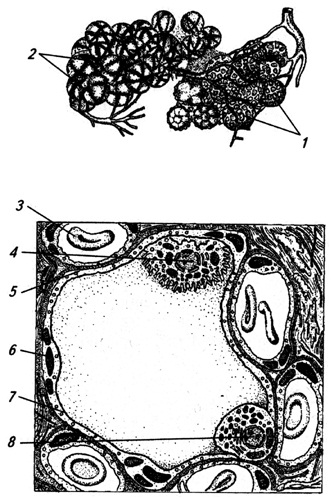
Small. 4.33. Budova alveoli walls:
1 - blood-bearing capillaries;
2 - bundles of elastic fibers;
3 - erythrocyte in the lumen of the capillary;
4 - alveolar macrophage,
5 - between pneumocytes;
6 - type I pneumatic cylinders;
7 - plavka surfactant;
8 - pneumocyl II type
Type I pneumocytes- strongly adhered cells, up to 0.2 microns in thickness, which fix the wall of the alveoli. Through the cytoplasm, the diffusion of gases occurs: acid and carbon dioxide. There are type II pneumocytes between the crimi cells. The stench is itself to fill the great secretory cells, which can be seen in the lumen of the alveoli.
The names of pneumocytes of both types are otochened by the basal membrane, as in bagatokh dilyankas they get angry with the basal membrane of the blood-bearing capillaries, alveolocapillary membrane.
Type II pneumocytes see the speeches of the transcendental nature, so that you can enter the warehouse surfactant. The rest is a foldable speech, which curls the inner surface of the alveoli and does not allow them to glue together in the daytime.
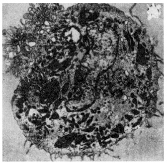
Legenevian macrophage. Most of the overwhelming cells in the intercalveolar walls and in the alveolar spaces are present in the presence of a large number of macrophages (Fig. 4.34.). The stench is established from the blood monocytes and goes through the alveolar wall into the lumen. The main function of the legene macrophages is the cutting of the saw and foreign particles from the alveolar holes.
Parasympathetic innervations of the fibers of the bloating nerve, stimulation of rapid smooth muscles of bronchioles.
razdratuvannya nice system, Navpaki, wiklikє її weakening.
Efficient nerve fibers are the most abundant for type II pneumocytes.
To get involved, in the legends of the afferent nerve fibers.
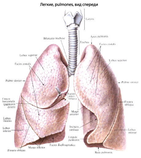
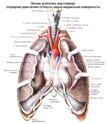
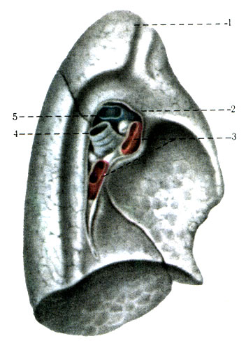
Pulmones, right and left, behind the shape are represented as two halves of a rasping cone. The right leg is shorter and wider than the left and the bigger the leg (Fig. 125). In the cutaneous lung, the base, basis pulmonis, becomes the lower diaphragmatic surface, the fades diaphragmatica, the upper part, apex pulmonis, is straightened up and stands 2-4 cm apart, the I ribs, and also two surfaces: the rib, rounded the costalis breast cancer, and medial, fades medialis, - I will be excluded. I will stay in the middle part, pars mediastinalis, turned up to the middle, and і ridge, pars vertebralis, lay down to the ridge. In the middle of the part, there is a heart-shaped impression, impressio cardiaca, which is especially relevant in the legend. On the same surface of the left legacy, one can see the clavicular boron and the boron aorta, and on the right - the boron stravohode. In addition, on the medial surface of both legends, the gates of the legends, hilus pulmonis, pass through them, the bronchi and the court pass through them, the roots of the lung, radix pulmonis.
The surface of the lung is interconnected by one side of the same two edges: the lower, margo inferior, and the lower surface of the medial and costal, and anterior, margo anterior, which is in front of the medial surface of the rib. Behind the place the transition of the medial surface into the rounded rib, and the edge there is a day.
Behind the help of interlobar gaps, fissurae interlobares, are easily subdivided into parts, lobi. The rights of the legend are divided into three parts: upper, lobus superior, middle, lobus medius, and lower, lobus inferior. The lower part of the additional oblique slit, fissura obliqua, is seen as a middle and upper part, like a horizontal slit, fissura horizontalis. In the legends, there are two parts: upper, lobus superior, and lower, lobus inferior, divided by an oblique split. On the lower half of the front edge of the legends, there is a heart virus, incisura cardiaca, pulmonis sinistri, in the lower part of the lung I set up a vistup - the tongue of the legends, lingula pulmonis sinistri, the late right middle parts.
The new generation of the legends expands with the establishment of the dichotomy. Until the end of the 1st fate of life, the number of legends grows 4 times, up to 8 rocks - 8 times, up to 12 rocks - 10 times. The top of the leg in newborns reaches the level of 1 rib, and the cordon passes through all the way, lower in the grown-ups.
Budova Legen
The legends are stored from the bronchial tubes, which fix the bronchial tree, and the collection of the legeny chickens, or the alveoli, which are located near the red bronchial tubes - the respiratory bronchial tubes.
Kintsevoy structural unit of the leg is acinus, which folds from the dychial bronchioles of the cytoplasm, which spreads to a number of alveolar passages and alveolar mice. 15-18 acinus put a legene part - a dilenca of legene fabric of a paramidal shape with a diameter of up to 1 cm (Fig. 126). Fragments vіdokremlenі one out of one with semi-woven cloths, in which veins and lymphatic judges pass. The legene part includes the bronchus (8-9th order of reddening), the legs of the leg artery, the bronchial artery, the nerves and the veins of the leg veins, the bronchial veins, the lymphatic judgments.
Large structural unit, macroscopically, є broncho-legene segment, segmentum bronchopulmonale, which is established in 2000-3000 small parts. Segments store parts of the lung in their own accord. The name is lightly covered with a legacy pleurora - a serous sheath, to avenge richly elastic fibers and covered with mesothelium.
The wall of the internal bronchial tubes is in the bud, lower in the head bronchi. In segmental bronchi and cartilage throats, they fall on the edge of the plate. The block of bone bronchioles is relieved by cartilage, the mucous ball is of strength, mucous clots are out of the way.
Segmental Budova Legen
In the lungs, there are 10 broncho-lenevic segments, such as a vascular segmental bronchus, a pulmonary artery, a bronchial artery and a vein, nerves and lymphatic judgment. Segments vidokreleleni one from one projectile with the resulting fabric, in which intersegmental legends pass through (Fig. 127)
Segments of the right legacy
Segments of the Legends
Analogous name may be segmental bronchi.
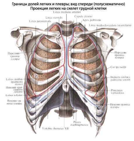
topography legend... It is easy to grow in the pleural cells (div. Rozdil Moche-stat system, viz.) Of the breast cell. The projection of the lungs onto the ribs of the warehouse of the lungs, as a person lives, begins to be visualized (percussion) and radiological. Develop a cordon of the upper lungs, front, back and lower boundaries.
The tops of the clavicle are 3-4 cm apart. The anterior border of the right side of the legend goes from the top to the second rib along the linea parasternalis and far along them to the sixth rib, the devil goes to the lower border. The front border of the left leg goes up to the third rib, so it’s the right one, and in the IV mid-ribbed edge, go horizontally to the linea medioclavicularis, the stars go down to the VI rib, fix the lower border.
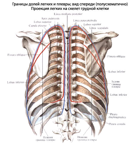
The lower boundary of the right leg runs gently in front of the cartilage of the VI rib backwards and down to the spinous ridge of the XI thoracic ridge, intertwining along the linea medioclavicularis the upper edge of the VII rib, along the linea axillaris media - the upper edge of the VIII rib, along the linea axillaris media - the upper edge of the VIII rib, along the linea axillaris media - the upper edge of the VIII rib - posterior linea scapularis - upper edge of X rib і along linea paravertebralis - XI rib. The lower boundary of the left leg is 1 - 1.5 cm lower than the right one.
The ribs on the surface of the lungs stick to the entire length of the chest wall, the diaphragmatic - to the diaphragm, the media - to the medial pleura and through it to the organs of the middle (right - to the right, unpaired arterial, lіvoї pіdclavicular artery, thoracic aorta, heart).
The topography of the elements of the root of the right and left lungs is not the same. At the roots of the right legacy, the right cephalic bronchus grows from above, below - the legene artery, in front and below which there is the legene vein. At the root of the left legacy, on the top lie the legene artery, behind and below the head bronchus, and below and in front of the bronchus, the leg veins grow.
X-ray anatomy of the lungs
on x-ray sign the breast cells are easy to appear in the eyes of the light, legendary fields, as they overwhelmed with oblique heavy shadows. It’s an intense time to grow up at the root of the legacy.
Sudden and nerves of the lungs
The lungs must lie down to two systems: 1) the vessels of a small stake, which can be used for gas exchange and transport of gases that have become blood-borne; 2) ships great stake blood circulation, so good food for the fabric of the leg.
Legendary arteries, which carry venous shelter from the right lumber, are distributed in legends on parts and segmental arteries and extending to the bottom of the bronchial tree. Establish a capillary netting around the alveoli, which will prevent the diffusion of gases into the roof, as well as with it. Constructed from the veins capillaries carry the arterial roof through the veins in the atrium.
Harchuvannya of the legacy tissue is seen through the bronchial arteries, and the venous blood - through the bronchial veins. However, offending the system and not being isolated will result in one kind of one - between the limbs of the bronchial and legenevius of the anastomoses.
Razrіznyayut glibokі and superficial lymphatic judges. Dzherelami gliboky vodina є lymphatic capillary fenestration near the endovian bronchioles, interatomic and interlobular prominences. The superficial vessels are formed from the pleural capillary fenestration. Lead the judges to go through the bronchi to the Legendary, Broncho-Legendary, broncho-tracheal and bifurkatsiynyh universities.
The innervations of the lungs are supported by plexus pulmonalis.
The lungs (pulmones) represent the head of the organization of the dichotomy, which can fill the entire chest empty, except for the middle. In the lungs, gas exchange is carried out, i.e., there is a clawing of the alveoli with blood rhytrocytes and the appearance of carbon dioxide in the alveoli, as the alveoli fall into carbon dioxide gas and water. In such a rank, in the legends, there is no way of describing twisted nobles, blood-bearing and lymphatic judges and nerves. Establishment of nobles for a meal and blood in a special room mental system quilting from the early stages of the embryonic and phylogenetic development. The insensibility of the acidic organisms lies at the stage of ventilation of the young children of the lungs, in addition to the ventilation and the fluidity of the blood flow, the density of the blood with hemoglobin, the fluidity of the diffusion of gases through the alveolar-capillary tissue of the springy membrane, the elasticity of the lungs I would like to bring one of these indicators to the destruction of the physiology of the situation and the possibility of the destruction of the functional destruction.
The name of the Budov legends is simpler to complete (Fig. 303). per form lighter nagadu cone, develops the top (apex), pіdstava (basis), ribbed surface (fades costalis), diaphragmatic surface (fades diaphragmatica) and medial surface (facies medians). Two remaining surfaces are excluded (Fig. 304). On the medial surface, the ridge part (pars vertebralis), the middle part (pars mediastinalis) and the heart in a vice (impressio cardiaca) are laid out. Liva glyboka is cordially depressed to complement the heart virizkoy (incisura cardiaca). In addition, the fades interlobares. The anterior edge (margo anterior) is visible, as well as the rib and medial surface, the lower edge (margo inferior) is seen on the rib and diaphragmatic surfaces. Lightly cover with a thin visceral sheet of pleura, through the yak you can see through the dark fabric of the fabric, which is located between the small pieces. On the medial surface, the visceral pleura does not curl the gates of the leg (hilus pulmonum), but descends below the viglyadi duplikatury under the name of the ligg. Pulmonalia.

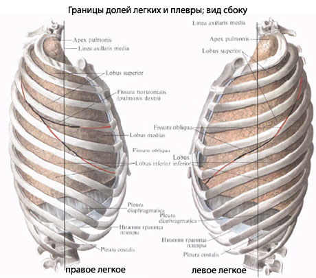
At the gates of the right leg, there is a bronchus, then the leg artery and vein (Fig. 304). At the top of the legends there is the Legend's artery, below the bronchus and the vein (Fig. 305). All the time to learn the roots of the legends (radix pulmonum). The root of the legend and the legend of the legend to settle the light in the singing position. On the costal surface of the right leg, a horizontal fissure (fissura horizontalis) and a lower braid (fissura obliqua) are visible. The horizontal slit is located between the linea axillaris media and the linea sternalis of the breasts and is located directly behind the IV ribs, and the braid of the slit is located directly behind the VI ribs. Behind, repaired from linea axillaris to linea vertebralis of the breasts, є one is furrowed, which represents an extended horizontal furrow. For the rakhunok, the cich is borozen in the right legend, the upper, middle and lower parts (lobi superior, medius et inferior) are developed. The most part is the lower, then the upper and the middle are the naymensha. In some legends, the upper and lower parts are seen, bordered by a horizontal slit. In the lower part of the heart, on the anterior edge is the testicle (lingula pulmonis). It is easier to lower the right one, which is tied to the lower positions of the left dome with diaphragms.
Mezhi Legen... The tops of the legs protrude 3-4 cm on the shoulder of the collarbone.
The lower boundary between the legs begins at the point of the ribs overretin with cleverly drawing lines on the breasts: along the linea parasternalis - the VI rib, along the linea medioclavicularis (mamillaris) - the VII rib, along the linea axillaris media - the VIII rib, along the linea scapularis - the X rib, along the linea scapularis - the X rib - at the head of the XI rib.
With the maximum inhalation, the lower edge of the legends, especially along the two remaining lines, descend by 5-7 cm. Naturally, between the visceral pleural layer, it sits behind the cordon of the lungs.
The front edge of the right and left lungs is designed on the front surface of the chest wall in a fashionable way. Having felt from the tops of the lungs, the edge should pass parallel to the edge of 1-1.5 cm one from one to the level of cartilage of the IV rib. At the end of the day, the edge of the left leg grows 4-5 cm, the cartilage of the IV-V ribs is not covered with light. The central impression (impressio cardiaca) is filled with the heart. The anterior edge of the lungs at the sternal end of the VI rib pass into the lower edge, the cordon of both lungs is zbigayut.
Internal structure of the lungs. lung tissue to be distributed on non-parenchymal and parenchymal components. Before the first one should lie down on the bronchial veins, on the legs of the leg artery and on the leg veins (except for the capillaries), the lymphatic judges and nerves, the semi-tissue projections, but to lie in the small parts, on the bronchial vessels of all the vessels The parenchymal part is stored from the alveoli - the alveolar bears and alveolar passages with the draining blood-bearing capillaries.
architecture of bronchial(Fig. 306). Right і lіviy legeny bronchi at the gate can be distributed to parts of the bronchi (bronchi lobares). All parts of the bronchi pass through the great throats of the Legendary artery, behind the vignette of the right upper lobe bronchus, which grows over the artery. Parts of the bronchi are divided into segmental ones, which last time in the form of an incorrect dichotomy up to the 13th order, ending in partly bronchial tubes (bronchus lobularis) with a diameter of close to 1 mm. Skin lesions have up to 500 parietal bronchial tubes. At the stage of all bronchial tubes, cartilage rings and spinal plates, reinforced with collagen and elastic fibers and are equipped with muscular elements. In the mucous membranes of the bronchial tree, there is a lot of growth in the mucous membranes (Fig. 307).

With the growth of the bronchus of the bronchus, the diagnosis is clearly new - bronchi terminates with a diameter of 0.3 mm, as well as the addition of the cartilaginous base and the viscosity of the single-ball prismatic epithelium. Kintsev bronchi, lastly expanded, form bronchioles of the 1st and 2nd order (bronchioli), in the walls of some kind of bladder spheres, the building of the bronchiole enlightens. The stench is added to the respiratory bronchioles of the 1st, 2nd and 3rd order (bronchioli respiratorii). Respiratory bronchioles are characterized by the appearance of appearance without the middle of the alveolar passages (Fig. 308). Respiratory bronchioles of the 3rd order appear with 15-18 alveolar passages (ductuli alveolares), the signs of which are fixed with alveolar bears (sacculi alveolares), but to replace the alveoli (alveoli). The system of nasal respiratory bronchioles of the 3rd order is stored in the acinus of the lung (Fig. 306).
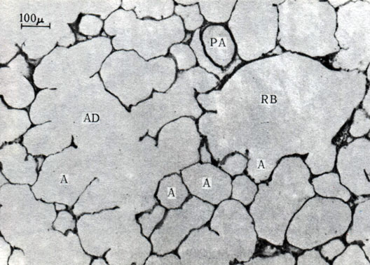
308. Histological development of the lung parenchyma of a young woman, which is stored without alveoli (A), which is often tied with an alveolar passage (AT) or respiratory bronchialitis (RB). RA - gilka of the legendary artery. × 90 (via Weibel)
budova alveoli... Yak bulo is designated as an alveoli, the alveoli enter the parenchyma warehouse and represent the end portion of the airway system, which develops gas exchange. Alveoli represent the type of alveolar passages and mice (Fig. 308). The smell may be in the base of a cone-like shape with eliptic peretin (Fig. 309). Alveoli fill up to 300 million; the stink is stored on the surface, equal to 70-80 m 2, ale dichal to the surface, i.e. emptying the alveoli into the roof and back. Alveoli pokriti malimi, great and vilny flat clays... Remaining also possible to phagocytose foreign particles. The cells grow on the basal membrane. The alveoli are cooled with blood-bearing capillaries, and the endothelial cells are stuck to the alveolar cells. Gas exchange is carried out at the same time. The endothelial-epithelial membrane is 3-4 microns.
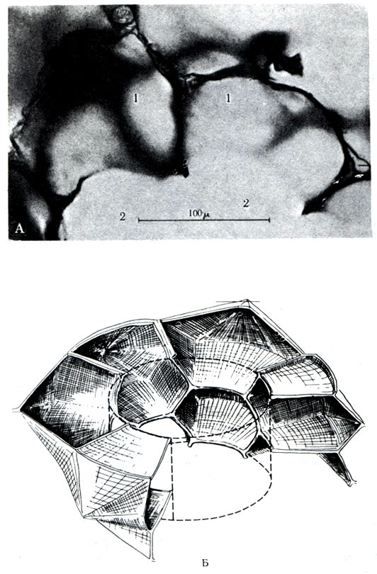
Between the basal membrane of the capillary and the basal membrane of the alveoli, there is a small interstitial zone, which is to remove elastic, collagen fibers and fibrils, macrophages and fibroblasts. Fibrous lightness gives a light fabric and elasticity; for her rakhunok i take care of an act of vidihu.
segments of the leg
Broncholegenic segments represent a part of the parenchyma, which includes the segmental bronchus and artery. On the periphery of the segment, one for one, and in contrast to the legacy, a small part of the clear prod- The skin segment has a cone-like shape, the top of the yak is turned to the top of the leg, and at the top - to the th surface. The intersegmental sticks have the leg veins. The cutaneous lung has 10 segments each (Fig. 310, 311, 312).


Segments of the right legacy
Segments of the upper part... 1. The upper segment (segmentum apicale) borrows the upper lung and the mass of intersegmental cordons: two on the medial and two on the costal surface of the lung, and the upper and the anterior, upper and posterior segments. The area of the segment on the costal surface is smaller, lower on the medial. Before the structural elements of the segment vorit (bronchus, artery and vein), the motile pidhid goes through the rosette of the visceral pleura in front of the vorit legends along the phrenic nerve. The segmental bronchus is 1-2 cm long, and it goes through the posterior trunk from the posterior segmental bronchus. On the thoracic plate, the lower boundary of the segment shows the lower edge of the II rib.
2. The posterior segment (segmentum posterius) grows dorsally to the upper segment and five intersegmental cordons: two - to be projected on the medial surface of the lung between the posterior and upper, posterior and upper segments of the lower triple part, to be seen back, back and front, back and upper segments of the lower part of the leg. The cordon, approved by the back and front segments, is arranged vertically and ends at the bottom on the fissura horizontalis і fissura obliqua stick. The cordon is located between the rear and upper segments of the lower part of the rear part of the fissura horizontalis. From the medial side to the bronchus, arteries and veins of the posterior segment, when the pleura on the posterior-upper surface rises from the side of the cob to the horizontal furrow. Segmental bronchus grows between arteries and veins. In the posterior segment, it becomes angry with the vein of the anterior segment and flows into the Legenev vein. On the surface of the chest wall, the posterior segment is designed between II and IV ribs.
3. The anterior segment (segmentum anterius) is located in the anterior part of the upper part of the right leg and there are five intersegmental cordons: two - pass on the medial surface of the lung, distribute the anterior and upper anterior segments of the medial three cordons pass along the costal surface between the anterior and upper, anterior and posterior, anterior, lateral and medial segments of the middle part. Artery of the anterior segment of the vein from the upper arm of the Legendary artery. The day of the segment by the tributary of the upper leg vein and rosette is larger than the segmental bronchus. The vertebral column of the segment can be ligated by placing the medial pleura in front of the lung. The segment will be welded to the edges of the II - IV ribs.
Segments of the middle part... 4. The lateral segment (segmentum laterale) on the side of the medial surface of the lung is projected only at the viglyad of the vuzkoy smog in an oblique interlobar furrow. Segmental bronchus rectifying backwards, to that loan segmentє back part the middle part is visible from the side of the costal surface. There are five intersegmental cordons: two - on the medial surface of the lateral and medial, lateral and anterior segments of the lower part (the remainder of the border of the medial segment of the lateral triangular furrow, the front between the horizontal furrow vertically from the middle of the horizontal furrow to the end of the oblique furrow, the other between the lateral and anterior segments and the vertical position of the horizontal furrow; the remainder of the inter-lateral segment sticks from the anterior and posterior segments of the lower part).
Segmental bronchus, artery and vein grow too quickly, until they can only go along the oblique boron below the lung. The segment is open to space on the chest plate between IV-VI ribs.
5. The medial segment (segmentum mediale) is visible on the costal and on the medial surfaces of the middle part. Winters of the intersegmental cordons: dvi - open the medial segment from the anterior segment of the upper part and the lower segment of the lower part. The front of the border is located with the front part of the horizontal furrow, the other - with the oblique furrow. On the costal surface there are also two intersegmental cordons. One line is repaired in the middle of the front part of the horizontal furrow and descend to the end part of the oblique furrow. The other border of the medial medial segment is from the anterior segment of the upper part and to stand behind the positions of the anterior horizontal furrow.
The segmental artery is seen from the lower arm of the Legendary artery. 4 segments at once from the artery. From it, the segmental bronchus grows, and then the vein is up to 1 cm. Access to the segmental lower muscle is lower than the lung through the interlobar sulcus. The cordon of the segment on the thoracic clitts is from the IV-VI ribs along the middle groin line.
Segments of the lower part... 6. The upper segment (segmentum superius) occupies the upper part of the lower part of the leg. The segment on the lines of the III-VII ribs is two intersegmental to the cordon: one between the upper segment of the lower part and the back segment of the upper part pass along the oblique boron, the other - between the upper and lower segments of the lower part. In addition to the importance between the upper and lower segments, it is necessary to intelligently advance the anterior part of the horizontal sulcus of the lung from the middle of the zlittya to the oblique sulcus.
The upper segment will remove the artery from the lower leg of the Legendary artery. The bronchus grows below the artery, and then the vein. The segment can be reached through the interlobar furrow through the spit. The visceral pleura is tinted from the side of the costal surface.
7. The medial basal segment (segmentum basale mediale) grows on the medial surface of the lower veins, sticks to the right anterior and lower empty veins; mak cordoni with anterior, lateral and posterior segments. Only 30% of all vapors are accepted.
The segmental artery is seen from the lower leg of the Legendary artery. Segmental bronchus є navischuyu gilkoyu LOWER LEFT bronchus; The vein grows lower than the bronchus and flows into the lower right Legeny vein.
8. The anterior basal segment (segmentum basale anterius) grows in the anterior part of the lower part. On the chest plate, the ribs VI-VIII appear along the middle groin line. There are three intersegmental cordons: the perch passes through the anterior and lateral segments of the middle part and forms an oblique interlobar furrow, the other - between the anterior and lateral segments; її projection on the medіїsurfaces її їѕ іѕ bіgаtіn with an ear of a legendary sound; the third boundary pass between the anterior and upper segments of the lower part.
The segmental artery spans the ear from the lower leg of the Legeny artery, the bronchus - from the side of the LOWER LEFT bronchus, the vein flows into the lower Legeny vein. The artery and bronchus can be promoted from the visceral pleurora at the bottom of the oblique interlobar sulcus, and the vein - from the lesions.
9. The lateral basal segment (segmentum basale laterale) is visible on the costal and diaphragmatic surfaces of the lung, between VII-IX ribs along the posterior groin line. There are three intersegmental cordons: persha - between the lateral and anterior segments, the other - on the medial surface between the lateral and medial, the third - between the lateral and posterior segments.
Segmental arteries and bronchus grow in oblique furrows at the bottom, and the vein - with a leg viscous ring.
10. The posterior basal segment (segmentum basale posterius) lies in the posterior part of the lower part, sticking to the ridge. Borrowing space between VII-X ribs. Seeing two intersegmental cordons: persha - between the posterior and lateral segments, the other - between the posterior and upper segments. Segmental artery, bronchus and vein grow in the glybin oblique furrow; it’s easier to get to them than an hour of operation goes from the medial surface of the lower part of the legacy.
Segments of the Legends
Segments of the upper part... 1. The upper segment (segmentum apicale) practically repeats the shape of the upper segment of the right leg. The appearance of the artery, bronchus and vein of the segment grows.
2. The posterior segment (segmentum posterius) (Fig. 310) with the lower boundary descend to the level of the V rib. The upper and posterior segment is often combined into one segment.
3. The anterior segment (segmentum anterius) is occupied by the very position, only the lower intersegmental boundaries pass horizontally along the III rib and the upper ovarian segment.
4. The upper lingual segment (segmentum linguale superius) grows on the medial and costal surfaces on the ribs of the III-V ribs in front and along the middle groin line and IV-VI ribs.
5. The lower ovarian segment (segmentum linguale inferius) is located below the anterior segment. The lower part of the intersegmental inter- segment is located behind the interlobar furrow. On the anterior edge of the lung, between the upper and lower ovarian segments, there is the center of the heart of the lung.
Segments of the lower part zbіgayutsya s the right legend.
6. Upper segment (segmentum superius).
7. Medial basal segment (segmentum basale mediale) non-perishable.
8. Anterior basal segment (segmentum basale anterius).
9. Lateral basal segment (segmentum basale laterale).
10. Posterior basal segment (segmentum basale posterius)
pleural beetles
The right and left of the pleural bears of the breast empty represent the older empty ones (celoma). The sides of the breast emptying are covered with the parietal leaf of the serous membrane - the pleura (pleura parietalis); from the parenchyma of the lung, the pleura visceralis pulmonalis grows. Between them, the empty pleura (cavum pleurae) is closed with an insignificant amount of ridini - close to 20 ml. Pleura is a large plan of Budovia, a serous membrane lures us, that is, the upper leaves, one to one, are covered with mesothelium, spread on the basal membrane and semi-tissue fibrous base from 3-4 balls.
The parietal pleura covers the walls of the breast, growing from f. endothoracica. In the area of the ribs of the pleura, it is easy to grow in the area. Falsely from the position of the parietal leaf, the rib, diaphragmatic and middle pleura are seen. The rest is planted from the pericardium and up to pass into the dome of the pleura (cupula pleurae), which hangs over the I rib by 3-4 cm, below go into the diaphragmatic pleura, from the front and back - into the rib, and through the bronchus, arteries and veins, there are three veins visceral leaf. The parietal leaf takes care of the fate of the three pleural sinuses: right and livy costo-diaphragmatic (sinus costodiaphragmatici dexter et sinister) and costo-mediastinal (sinus costomediastinalis). The right-hander and the left-handed person are first moved out of the dome with diaphragms and between the costal and diaphragmatic pleura. Costomediastinal sinus (sinus costomediastinalis) is unpaired; The cisterns represent a reserve place of the pleural empty space, where the tissue should enter for an hour. In pathological processes, if there is shelter in the pleural bears, there is a stench in the perch, and the stench accumulates in the sinuses. Adhesions yak inherited pleural effusions in front of them in the pleural sinuses.
Between the parietal pleura
The parietal pleura occupies a large area, lower visceral. Liva pleural emptying is more and more right. The parietal pleura grows up to the head of the first rib and the illumination of the pleural dome (cupula pleurae) protrudes 3-4 cm above the first rib. Backward, the parietal leaf descends to the head of the XII rib, de pass into the diaphragmatic pleura; ahead to right side Having spread out from the capsule of the sternoclavicular corner, it descends to the VI rib along the inner surface of the sternum, passing into the diaphragmatic pleura. The malignant parietal leaf goes parallel to the right pleura to the cartilage of the IV rib, then extends to the left by 3-5 cm and on the rivni VI rib passes into the diaphragmatic pleura. The pericardial tricut, not close to the pleurora, growth up to IV-VI ribs (Fig. 313). The lower boundary of the parietal leaf begins at the center of the cleft of the upper lines of the breasts and ribs: along the linea parasternal - the lower edge of the VI rib, along the linea medioclavicularis - the lower edge of the VII rib, along the linea axillaris media - X paratebral, along the linea scapularis - XIver - to the lower edge of the body of the XII chest ridge.

Vіkovі singularities і pleurisy
At the newly born, the upper part of the light less, Nіzh at the child until the end of the first fate of life. Before the period of the statutory dosage it is easier in the case of the lungs of a newly born child to grow in volume 20 times. The right legacy to develop more intensively. In a newborn, in the walls of the alveoli, there are few elastic fibers and a lot of fluffy fabrics, which is indicated on the elasticity of legends and quick development of the filling during pathological processes. In particular, those who, in the first 5 years of life, increase the number of alveoli and the order of bronchial discharge. Acinus tilka in a child of a 7-rider viku nagadu for a grown-up budova acinus. Segmental Budova is clearly twisted in the course of life. After 35-40 years of age, there will be revolutionary changes that govern all tissues of their organs. The epithelium of the dysfunctional nobles grows thin, the elastic and reticular fibers are stretched out and fragmented, to replace the low-loss collagen fibers, infusion of pneumosclerosis.
In the pleural sheets of the lungs, up to 7 rocks are parallel to the increase in the number of elastic fibers, and the bagatosharovaya mesothelium pleurisy changes to one ball.
mechanizm dikhannya
The parenchyma of legends is to take revenge on the elastic fabric, as it is good to go for the expansion of the original volume. To that Legeneva, the dichotomy is possible in that vipad, as if the grip in the turn-over paths will be more, there will be no calls. Rise of a rotary vise from 8 to 15 mm Hg. Art. dolaє opіr elastic tissue parenchyma of the lungs. It should be seen when the breast tissue is enlarged during the period of inhalation, if the parietal pleural leaf changes at once with the diaphragm and the ribs, which leads to the increase in pleural beads. The visceral leaf slides passively behind the paralytic under the grip of the fluid stream in the pleural cells and lungs. It is easier to be in hermetically sealed pleural bears, in the stage of inhaling all the intestines. In the stage of the vidihu of the breast tissue, it relaxes and the parietal leaf of the pleurisy at the same time with the breast tissue approaches the center of the breast emptying. Legeneva fabrics, due to elasticity, change in volume and vishtovkhuy.
In quiet cases, if there are abundant collagen fibers (pneumosclerosis) in the tissue, and the elastic traction of the lungs is destroyed, such difficulties can lead to the expansion of the legends (emphysema) and the breakdown of gas exchange (hypoxia).
When a parietal abnormally visceral pleural leaf is damaged, the hermeticity of the pleural empty space breaks down and pneumothorax develops. In general, it is easier to subside and to imitate the mental function. In case of a defect in the pleura and defect in the pleura, it is easier to get involved in the reaction.
For an hour, I breathe in the dome with diaphragms to lower by 3-4 cm and the spine-like ribs of the front end go forward and up the hill. Newborns and children of the first rocky life have a dichotomy behind the rakhunok with diaphragms, so as the ribs do not dim curvature.
In case of a calm dichanna, you can drink and vidih in a warehouse of 500 ml. The central part of the lungs is stored in the lower part of the lungs. It is practical not to take part in the gas exchange in the upper leg. With a quiet dichan, the part of the alveoli becomes covered with a closure of the fast-flowing ball of the respiratory bronchioles of the 2nd and 3rd order. Only in case of physical robots and a high degree of energy, the entire legene tissue is included in the gas exchange. Life The number of legends for choloviks should be 4-5.5 liters, for women - 3.5-4 liters, and they should be stored for a quiet, supplementary and reserve watch. For the maximum vidihu in the legends, 1000-1500 ml of surplus drink should be consumed. In case of calm dichannia, add 500 ml of alcohol (dichal dizziness). Dodatkovy povitrya in the general population of 1500-1800 ml to take place at the maximum inhalation. A reserve drink in the general population of 1500-1800 ml is transferred from the lungs in the presence of a vidih.
Diseased ruins are reflexively detected 16-20 times per quill, the frequency of dysfunction is rather small and high. For an hour I breathe, if the grip falls into the pleural emptying, the rush of venous blood rises to the heart and the lymph rises from the thoracic duct. In such a rank, glyboke dikhannya is beneficially infused into the blood circulation.
roentgenograms legeniv
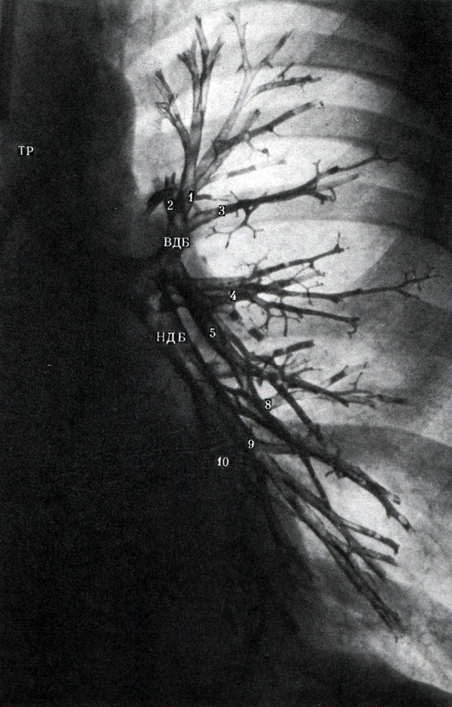
When X-ray diffraction, it is easy to see oglyadov, straight and innocent, and also targeted by radiographs and tomography until the end of the day. In addition, it is possible to vivify the bronchial tree, resembling the bronchi with contrasting words (bronchogram).
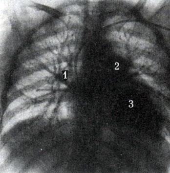
On the lookout in the front projection, you can see the organization of the breast emptying, chest wall, Diaphragm and small stove. On the roentgenogram, one can see the right (more) or more (less) of the field, at the bottom, surrounded by a furnace, in the middle - with a heart and aorta. Legendary fields are formed with a clear tone of blood-bearing blood-bearing vessels, well contoured on the light aphids, made with full-tissue prods and alveoli and other bronchial tubes. Tom, on one їkh obsyagu fall even more richly woven fabrics. Legenevian babies on aphids of Legenevian fields are built up from short bunches, gurts, specks, which may turn out to be contoured. Tsei Legenevii babies know that it is easier to draw lightness into the result of the build up, or the loss of legenevo tissue (atelectasis); when the legacy fabric is ruined, more lights are used. Between parts, segments, parts in the norm are not visible.
Greater intense lightness is spared to the norm for the rakhunok of more great Sudins. Evil is the root of the lung at the bottom of the bottom of the heart, and at the top is the heart and the wide is the legendary artery. right tin root of the lung mensch is contrasting. Between the heart and the right legacy artery є light from the industrial and LOWER LEFT bronchial tubes. The right dome with diaphragms grows on the VI-VII ribs (in the phase of inhalation) and in the first place, below the edges. Before we rule to lie down intensively, the stove is intense, before we lie down - turning a little bit to the scrubber.
On an oblique X-ray in a normal projection, it is possible not only to look in greater detail on the Legendary field, but to project the Legendary segments, which, in general, cannot be projected one by one. On the whole sign it is possible to establish the scheme of segment rosetting. On a normal basis, depending on the intensity as a result of the superposition of the right and left lungs, the structure of the nearest lung is more clearly rotated. At the upper part of the sign, you can see the upper part of the lung, on which partly one can see the back and the belts of the upper part from the thin front border: at the bottom you can see the cover of the dome with diaphragms, so you can fit the ribs of the guest - the ribs in the middle of the ribs ribs and scapula. The Legenevian field is divided into two larger dylyankas: the posterior sternum, surrounded by the sternum, the heart and the aorta, and the posteriorly, which is located between the heart and the ridge.
The trachea can be seen at the viglyadi of the light brown to the level of the V chest ridge.
A targeted X-ray image will add additional signs, details are found in the most beautiful images, and are often stagnant when diagnosing problems pathological changes in the upper legs, costal-diaphragmatic sinuses, lower for the appearance of normal structures.
Tomograms (pozharovі signs) are especially effective for implantation of the lungs, as in this case, a ball can be seen on the image, so that it lies on the lobule of the lung.
On the bronchogram, the bronchial tubes are filled with a contrasting speech, it is inserted through a catheter into the head, partial, segmental and lobular bronchi, and the stitch of the bronchial tree is inserted. Normal bronchi may show some clear contours, after the last few days in diameter. Contrasting bronchi is clearly visible on the ribs and the root of the leg. With inhalation, normal bronchi podovzhuyut and expand, with vidihu - navpaki.
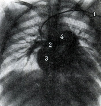
On a direct angiogram a. pulmonalis is 3 cm in diameter, 2-3 cm in diameter, and is located on the middle of the ridge on the level of the VI thoracic ridge. Here won’t go on the right and left of the head. It is also possible to differentiate all segmental arteries. In the upper and middle part, go into the upper Legenev vein, which can be positioned, and the veins of the lower part - into the lower Legenew vein, rosetted horizontally along the line to the heart (Fig. 314, 315).
phylogenesis of the lungs
In aquatic creatures there is a chickberry apparatus, and a pharynx squash is thin. Chaffinches develop in all the ridges, and in the terrestrial stench there is a lichen in the membranous period (div. Development of the skull). In addition to the chickberry apparatus, to the organs of the chick, the superficial and labyrinth apparatus, which represent the loss of the pharynx, should lie on the back of the child. Bagato ribi dodatkovo up to the finchs under their mayut gut dikhannya. When kovtannі povіtrya blood-bearing intestine, soak the muslin. The amphibians of the school also have the function of a supplementary organ of dichotomy. Before the additional organs are introduced a swimming mikhur, which is obtained from the stravohod. It is easy to walk around the pair of large-chamber swimming gears, similar to those that are used in double-running and ganoid ribs. Tsі bulbashki, so yak lungs, will get the blood of 4 chiffchal arteries. In such a rank, the swimming michur is a combination of the pre-organ dyhannya in aquatic creatures, which is transformed into the main organ of dikhannya in terrestrial ones.
The evolution of the lungs and polygons in that, in a simple microscope, there are numerical partitions and empty spaces for the improvement of the vessel and the epithelial surface, as well as contact with the witnesses. Lungs emerged in 1974 from the largest Amazon ribi arapaima, which is strictly pulmonary. She has a chickberry so she only has 9 days of life. Spongy lungs tied with blood-bearing vessels and cardinal tail veins. The blood from the lungs comes to the great left posterior cardinal vein. The valve of the pechinkovo vein regulates the blood flow in such a rank that the heart becomes sick with arterial blood.
Tsі danі to signal about those that in the lower water creatures є all transitional forms from the water reaction to the terrestrial one: dumb bears, Lungs. In amphibians, plasuns, lightly rozvineni are rotten, as there are a small number of alveoli.
In birds of light, small-sized and lie on the dorsal part of the sternum, do not cover the pleurora. The bronchi will grow out of the wiggle bears, which are overwhelmed by the shkіroy. For an hour, the birds are encouraged to play with the birds. The majority of the lungs of birds from the lungs of the pilgrims are those that do not end up blindly, like those of the birds, with the alveoli, but the anastomoses with the wind capillaries.
In all savts, in the lesions, the bronchial tubes, which are obtained from the alveoli, develop beforehand. Only alveolar passages represent a surplus of light empty amphibians and plasuns. In ssavts, small parts of the segments, in the lungs, the central dikhal way and alveolar part. The alveoli are especially significant. For example, the area of the alveoli near the cat is 7 m 2, and the area of the horse is 500 m 2.
pulmonary embryogenesis
The bookmark is easy to repair from the illumination of the alveolar bear from the ventral wall of the stravohode, covered with a cylindrical epithelium. On the 4th type of the embryonic development in the right legend, there are three bears, in the left - two. Navkolishnє mesenchyme mice form the connective tissue base and bronchi, kudi grow blood-bearing judges. The pleura is vinic from the somatopleuria and splanchnopleuria, which is a second empty of the embryo.


