The central part of the frontal part. Zvivini to the brain
At the anterior viddil of the skin lesion of the brain, there is a loboval part, lobus frontalis. It ends in front of the frontal pole and is bordered at the bottom by the lateral furrow, sulcus lateralis (Sylvian boron), and behind - gliboka central furrow(Fig. 124, 125). The central beard, sulcus centralis (Roland's beard), roasted at the frontal area. You have to repair at the upper part of the medial surface of the glass great brain, Rozsіkє yogo upper edge, go down, do not digest, on upper lateral surface go down and end, the trochies do not reach lateral furrow... In front of the central furrow, and parallel to it, the precentral furrow, sulcus precentralis, grows. It remains to end at the bottom, not reaching the lateral furrow. The precentral furrow is not easy to digest in the middle part and to be stored in two independent furrows. From the precentral furrow, the upper and lower forehead furrows, suici frontales superior et inferior, are directed forward. The stench of rosetting may be parallel one to one and extend the upper lateral surface of the frontal part on the zivini. Between the central sulcus back and the precentral sulcus in front of central zivina, Gyrus precentralis (anterior). The upper frontal furrow lies on the upper frontal part, gyrus frontalis superior, which occupies the upper part of the frontal part. Between the upper and lower frontal furrows, the middle of the frontal zvivin, gyrus frontalis medius, is removed. The bottom of the lower frontal furrow is the lower frontal zivin, gyrus frontalis inferior. At the bottom of the zvivin, there are branches of the lateral furrow protruding from the bottom: the upper head, ramus ascendens, and the front head, ramus anterior. The lower part of the frontal part, which hangs over the anterior part of the lateral furrow, into three parts. Pokrishechnaya part (lobova pokrishka), pars opercularis (operculum frontale), roztashovani between the viscous and the lower side of the precentral furrow. The tsia part of the frontal part has given it this name to the fact that it looks like an island part (island) to lie in the groove of the furrow. The triangular part, pars triangularis, is located between the viscous back and front side. The orbital part, pars orbitalis, lies downward towards the frontal head, extending onto the lower surface of the frontal part. In a whole muscle, the lateral furrow expands, in conjunction with which they call the lateral fossa of the great brain, fossa. lateralis (cerebraiis).
lobova part... At the posterior end of the last surface of the part, the sulcus precentralis pass parallel to the sulcus centralis. There are two furrows in the lateral direction: sulcus frontalis superior et sulcus frontalis inferior. The staff of the lobova part will be divided into chotiri zivini - one vertical and three horizontal. Vertical zivina, gyrus precentralis, is found between sulcus centralis and sulcus precentralis.
Horizontal sections of the frontal part offensive:
1) upper lobova, gyrus frontalis superior, Yaka ide vishche sulcus frontalis superior, Parallel to the upper edge of the pivkule, medial surface;
2) middle lobov zvivin, gyrus frontalis medius, Stretch against the upper and lower frontal furrows and
3) lower lobova zvivina, gyrus frontalis inferior, Accommodation mіzh s ulcus frontalis inferiorі lateral furrow.
The lateral furrows, which enter into the lower forehead, move to the three parts: pars opercularis, Scho to lie with the lower end sulcus precentralisі ramus ascendens sulci lateralis, pars triangularis, Which is located between the lumps of the lateral furrow, i, nareshty, pars orbitalis, Scho take revenge in front of ramus anterior sulci lateralis.
Lobova part of the city central boron, sulcus centralis... You should fix on the medial surface of the brain, go to the upper-lateral surface, where along the trochus obliquely, backwards, and not to reach the lateral groove of the brain (div. Fig. ,,,).
Approximately parallel to the central boron grow precentral boron, sulcus precentralis, Ale won not reach the upper edge of the pivkule. Predcentral sulcus well ahead precentral zivin, gyrus precentralis.
Upper and lower forehead furrows, sulci frontales superior et inferior, Head towards the precentral furrow forward. Stink the frontal part of the upper lobovu zivin, gyrus frontalis superior, Yaka roztashovana in the upper frontal furrow and prostrate on the medial surface of the pavcule; middle lobovy zivin, gyrus frontalis medius, Yaku round up the upper and lower forehead furrows. The orbital segment of the zivini goes over to the lower surface of the frontal part. At the front end of the middle frontal zivini develop the upper and lower parts. Lower lobova zvivina, gyrus frontalis inferior, Lying between the lower frontal furrow and lateral furrow brain and the lateral groove of the brain extending into a number of parts (div. Dal).
Lateral beard, sulcus lateralis, - one of the most common sulci in the brain. There is a small part of the Kremlin from the front and the time. Zalyaga lateral furrowed on the upper-lateral surface of the cutaneous eruption and going from top to bottom and forward. In the depths of the furrows, perishing will grow - lateral fossa of the great brain, fossa lateralis cerebri The bottom is like a call to the surface of the island.
From the lateral furrows of the mountain, enter other furrows, called the heads. Naybilsh post it from them є vishidna gilka, ramus ascendens, і front head, ramus anterior; the upper-posterior furrow is called back gilkoyu, ramus posterior(Div. Fig.,).
The lower lobovy zivina, in the boundaries of which there is a hanging and a front head, is divided into three parts (div. Fig.): Rear - pokrishechno chastina, pars opercularis, I will be interlaced in front of the vishіdnіy gіlkoyu; middle - triangularis, pars triangularis To lie between the front and front legs, and the front - orbital part, pars orbitalis, Roztashovanu between the horizontal head and the lower lateral edge of the frontal part.
tim'yana chastka(Div. Fig. ,,,) Zalyaga backwards from the central furrow, like from the frontal part. from early part the part of the tim'yana is separated by the lateral sulcus, from tiled parts- in part tim'yano-potilichnoy furrows, sulcus parietooccipitalis.
Parallel to the precentral zviviny pass postcentral zivina, gyrus postcentralis, Beaten in the back postcentral furrow, sulcus postcentralis... From her backward, parallel to the later slit of the great brain, where intraparietalis, sulcus intraparietalis To extend the back-upper side of this part of the area into two sections: upper tim'yanu part, lobulus parietalis superior, What lies in the intra-parietal sulcus, i lower tim'yanu part, lobulus parietalis inferior, Roztashovanu to the bottom of the intra-parietal furrow. At the bottom of the time, there are two small differences: supra-marginal zivina, gyrus supramarginalis Lie anteriorly and locks the rear outward or lateral furrow, and locks backward toward the front kutovu zivin, gyrus angularis, Yaka zamikaє the upper edge of the furrow.
Between the viscid and the posterior groove of the lateral groove, a measles dilenka is removed from the brain, so that a yak is recognized fronto-tim'yana pokrishka, operculum frontoparietale... It includes the rear part of the lower frontal part, the lower part of the precentral and postcentral part and the lower part of the front part of the temporal part.
Potilychna part(Divine Fig. ,,,) There are not many cordons on the opaque surface, but it seems to be on top of the time and early part, behind the vignette of the upper end of the temporal medical struggle, as it often grows on the surface of 'yanoi. All three superficial pylitic parts: the tumor is lateral, the median is flat and the lower one is curved, it is rounded to the outline of the moss, there are a number of furrows and the zvivin.
Furrows and growths on the lateral surface of the pylitic part are not permanent and are often not the same in both sprouts.
Nybilsha s borozen - transverse potilichna boron, sulcus occipitalis transversus... In the case of an extension of the posterior intra-parietal sulcus and in the posterior viddili go into non-posterior lunate furrow, sulcus lunatus.
Approximately 5 cm to the front of the pole of the tilting part at the lower edge of the upper lateral surface of the pivot є loss - presealing virizka, incisura preoccipitalis.
skroneva part(Div. Fig. ,,) Maє naybilsh pivot to the cordon. At them, they develop a puffy lateral surface and a lower one. The blunt pole of the early part of the furrows is forward and downward. Laterally, the great brain is furrowed with a sharp edge towards the frontal part.
Two furrows, rots on the upper lateral surface: upper skroneva borozna, sulcus temporalis superior, і lower skroneva borozna, sulcus temporalis inferior, Sliding parallel to the lateral to the brain, subtract the part by three edges of the brain: upper, middle і lower, gyri temporales superior, medius et inferior.
These delinkas of the early part, which have their own outer surface, are straightened in the lateral groove of the brain, porous by short transverse skronevny borozy, sulci temporales transversi... Mіzh tsimi borozyayut 2-3 short transverse skronevі zivini, gyri temporales transversi, Po'yazany with zviviny of the early part and an island.
Ostrivkova part(Ostrіvets) (div. Fig.) lateral fossa of the great brain, fossa lateralis cerebri.
Vona is a three-sided pyramid, brutalized with its apex - the pole of the island - forward and named, towards the lateral furrow. From the periphery of the ridge of the frontal, temporal and early parts, which take part in the establishment of the stems of the lateral furrow of the brain.
Pidstava island with three sides otochen circular furrowed island, sulcus circularis insulae, Yaka at the bottom surface of the island step by step znika. There is not a lot of growth for a whole lot of people - porig island, limen insulae To lie on the cordon with the lower surface of the brain, between the ostrich and the anterior speech.
The surface of the island is prone to gliboka central furrow island, sulcus centralis insulae... Tsia boroznaya razdіlyaє ostіvets on the front, large, and rear, mensha, parts.
On the surface of the island, the number of others is larger zivin island, gyri insulae... The front part of the ma kіlka short zvivin island, gyri breves insulae, Rear - often one dovgu zvivin island, gyrus longus insulae.
furrows i Zvivini to the brain: upper-lateral surface
[ sulci and gyri of the brain mantle: the superlateral surface ]
- Bock C.E. (1809-1874). Handbuch der Anatomie des Menschen. Leipzig, 1841.
Translated by: Ronald A. Bergman, PhD; Adel K. Afifi, MD, MS; Julie L. Bates, BSS; The University of Iowa.
Atlas of Human Anatomy.
Karl Ernest Bock (1809-1874). Atlas of human anatomy.
Shifting by authoritative fakhivtsy on English mov classic curriculum, prepared with good accuracy and precision. Recommended to be victorious in the modern day.
= Access to the reference.
URL: http://www.anatomyatlases.org/atlasofanatomy/index.shtml quotation- Gray H., (1821-1865). The Digestive Apparatus. In: Henry Gray. Anatomy of the Human Body, (1918).
Henry Gray (1821-1865). Herbal apparatus. In the book: Anatomy of tila people.
Really broken up and kindly illustrated. Recommended to be victorious in the modern day.
= Access to the reference.
URL: http://www.bartleby.com/107 quotation- Clack J.W.
Human Anatomy.
Compilation: 1. Human Anatomy, F. H. Martini et al. 2003 4th ed. 2. The Coloring Review Guide to Human Anatomy, H. McMurtrie & J.K. Rikel, 1990. 3. Human Anatomy Lecture Outlines, James W. Clack, 9th ed., 2004.
Anatomy of a human.
Compilation of three children. Wonderful illustrations.
= Access to the reference.
URL: http://iupucbio2.iupui.edu/anatomy quotation- University of Wisconsin Medical School. ...
In: Global Anatomy. University of Wisconsin Medical School.
University of Neurology Resources. In materials: Zagalna anatomy.
= Access to the reference.
URL: http://www.medsch.wisc.edu/anatomy/anatomy.htm quotation- Terence H. Williams, M. D., Ph.D., D. Sc.; Nedzad Gluhbegovic, M. D., Ph.D .; Jean Y. Jew, M.D.
The Human Brain: Dissections of the Real Brain. In: Virtual Hospital of The University of Iowa.
Human mozok: Peretini of real drugs.
Really broken up and kindly іlustrovane is the boss.
= Access to the reference.
URL: http://www.vh.org/adult/provider/anatomy/BrainAnatomy/BrainAnatomy.html. quotation- Brain Sections. Electronic Atlas. In: Teaching Materials of the Department of Neurobiology and Pharmacology of the Northeastern Ohio Universities College of Medicine.
Electronic Atlas: Peretini Moses.
= Access to the reference.
URL: http://riker.neoucom.edu/DEPTS/NEUR/WEB/atlas/index.htm quotation- The Partners in Assistive Technology Training and Service (PATTS) program. Caldwell Community College and Technical Institute. Nervous System: CNS and PNS. In: PATTS Anatomy.
Nervous system: CNS and PNS. At the core: PATTS Anatomy.
Really broken up and kindly іlustrovane is the boss.
= Access to the reference.
URL: http://webschoolsolutions.com/patts/systems/anatomy.htm quotation- John Mazziotta, MD, PhD; Arthur Toga, PhD; Alan Evans, PhD; Peter Fox, MD; Jack Lancaster, PhD; Karl Zilles, MD, PhD; Roger Woods, MD; Tomas Paus, Md, Phd; Gregory Simpson, Phd; Bruce Pike, phd; Colin Holmes, Phd; Louis Collins, PhD, Paul Thompson, PhD; David Macdonald, PhD; Marco Iacoboni, MD, PhD; Thorsten Schormann, PhD; Katrin Amunts, MD; Nicola Palomero-Gallagher, PhD; Stefan Geyer, MD; Larry Parsons, PhD; Katherine Narr; Noor Kabani, PhD; Georges le Goualher, PhD; Jordan Feidler; Kenneth Smith, PhD, Dorret Boomsma, PhD, Hilleke Hulshoff Pol, PhD; Tyrone Cannon, PhD; Ryuta Kawashima, MD, PhD; Bernard Mazoyer, MD, PhD. A Four-Dimensional Probabilistic Atlas of Central Nervous System.
Chotirivimirny imovirnisny atlas to the brain of the people.
Describe the atlas.
= Access to the reference.
URL: http://www.mitre.org/work/best_papers/best_papers_01/mazziotta_atlas/mazziotta_atlas.pdf. quotation- Chris Rorden. Neuroanatomy Atlas.
Neuroanatomy: atlas.
Really broken up and kindly іlustrovane is the boss.
= Access to the reference.
URL: http://www.psychology.nottingham.ac.uk/staff/cr1/anatomy/home.html quotation- CALnet Programs for Anatomical Science Students: Neuroanatomy.
Neuroanatomy.
= Access to the reference.
URL: http://137.222.110.150/calnet/Introanat/Introanat.htm quotation - Gray H., (1821-1865). The Digestive Apparatus. In: Henry Gray. Anatomy of the Human Body, (1918).
- Bergman R.A., Afifi A.K., Heidger P.M. Section 17. Central Nervous System. In: Atlas of Microscopic Anatomy: A Functional Approach: Companion to Histology and Neuroanatomy: Second Edition. The Virtual Hospital. The University of Iowa.
Central nervous system. At the core: Ronald A. Bergman, Adel K. Afif, Paul M. Haidger: “Atlas of Microscopic Anatomy. Functional pidhid ".
Dozens of high-quality images of diagnostic histological preparations and descriptions. Look around.
= Access to the reference.
URL: http://www.anatomyatlases.org/MicroscopicAnatomy/MicroscopicAnatomy.shtml. quotation- Nervous Tissue. In: Histology Atlas. The University of Wisconsin Medical School. Department of Anatomy. John K. Harting, PhD, Chair.
nerve tissue
Dozens of high-quality images of diagnostic histological preparations with descriptions and without descriptions (in vibration).
= Access to the reference.
URL: http://www.medsch.wisc.edu/anatomy/histo/htm/ttoc.htm. quotation- Nervous System. In: The HistoWeb. The University of Kansas Medical Center.
Nervous system. At the Kerіvnistvі: "Histological Atlas".
Dozens of high-quality images of diagnostic histological preparations. Inventories.
= Access to the reference.
URL: http://www.kumc.edu/instruction/medicine/anatomy/histoweb/. quotation- John A. McNulty, Ph.D. Nervous Tissue. In: Histology. Function of the Human Body Lessons. Loyola University Chicago Stritch School of Medicine.
Nerve tissue. At the kerіvnistvі: "Histology".
Dozens of high-quality images of diagnostic histological preparations and descriptions.
= Access to the reference.
URL: http://www.meddean.luc.edu/lumen/meded/Histo/frames/histo_frames.html. quotation- William A. Beresford, MA, Ph.D. Central Nervous System. In: William A. Beresford, MA, Ph.D. Histology Full-Text. The Anatomy Department, West Virginia University.
Central nervous system. In the book: "Histology".
Pidruchnik of histology.
= Access to the reference.
URL: ../../Athens/Academy/1575/. quotation- Linda Dalton Nervous Tissue. In: Histology Lab Review Carousels. The University of Texas - Houston Medical School.
Nerve tissue. At the navchalny messenger "Physiology of people. Inspection of laboratory robots ".
Dozens of high-quality images of diagnostic histological preparations. We are informed about those.
= Access to the reference.
URL: http://medic.med.uth.tmc.edu/edprog/histolog/carousel.htm. quotation - Nervous Tissue. In: Histology Atlas. The University of Wisconsin Medical School. Department of Anatomy. John K. Harting, PhD, Chair.
anatomy
Histology
physiology
The human nervous system. In the case: "Stories of the biology of Dr. Kimbol"
Really broken up and kindly іlustrovane is the boss.
= Access to the reference.
URL: http://www.ultranet.com/~jkimball/BiologyPages/ quotation
Nervous system. At the biology certificate.
Really broken up and kindly іlustrovane is the boss.
= Access to the reference.
URL: http://ridge.icu.ac.jp/biobk/biobooktoc.html quotation
Neurons and nervous system (part I). Neurons and nervous system (part II). "Physiology of People". Music notes before lectures.
Really broken up and kindly illustrated initial materials.
= Access to the reference.
URL: http://www.biology.eku.edu/ritchiso/301syl.htm quotation
Rukhovi structures of the spinal cord. At the beginning of the book: Clinical neurophysiology. A short, good illustration of photographs and diagrams..
quotation
URL: http://thalamus.wustl.edu/course
Cellular Properties Database (CellPropDB). The repository for data regarding membrane channels, receptor and neurotransmitters that are expressed in specific types of cells. The database is presently focused on neurons but will eventually include other cell types, such as glia, muscle, and gland cells.
Danikh base "Neuron". Information about neurons and cells with strong smells together. Data about the channels of membranes, neurotransmitters for neurons, glial, muscular cells, and cells of zalosi. Change materials. Nauchni diagrams. posilannya.
quotation
URL: http://senselab.med.yale.edu/
Biological psychology.
Really broken up and kindly іlustrovane is the boss.
= Access to the reference.
URL: http://psych.athabascau.ca/html/Psych402/Biotutorials/ quotation
Mozok and behavior
Really broken up and kindly illustrated initial materials.
= Access to the reference.
URL: http://courses.umassmed.edu/mbb1/2003/index.cfm quotation
Zbori resources
Really broken up and kindly illustrated initial materials.
= Access to the reference.
URL: http://faculty.washington.edu/chudler/neurok.html quotation
In: Eric H. Chudler, Ph.D.
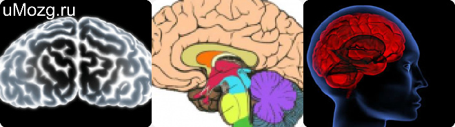
The bark of pivkul is covered with furrows and zivini. Among them, the most highly lying, firstly established furrows, are developed, which are divided into parts of the brain. Sylvieva boroznaya to the Kremlin part of the frontal area from the skernevoy dilyanka, Rolandova є to the cordon between the frontal and temporal parts.
Boroza of the temporal region grows on the medial area of the cerebral podvkuli and extends the potency of the day with the timyanim. The upper-lateral area is not similar to the cordon and cannot be divided into parts.
The median area can be found on its own lumbar furrow, as it passes into the furrow of the hipocampus, at the same time the median brain, signs for displaying the function of scent, from the other parts.
The furrows of the secondary designation for their budova, in the context of the primary ones, are designated for the distribution of parcels on a part - zivini, which are roasted from the zvnishnaya part of a similar type of zivin.
I am developing the third type of borozen - third, or as they call it - nameless. The stench is used to give a specific form of measles, as well as to increase the surface area of measles.
On the glybin, in the lower part of the biches, a part of the island has been removed. It was sharpened from the sides with a circular furrow, and the whole area is permeated with folds and hollows. For their functions, there is a sense of knowledge with the brain of scent.
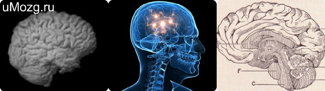
Talk about the brain, I want to go to my brain and look at it Anatomy Budova More details.
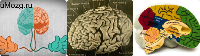
Otzhe, dermal injection has three types of surface: medial, lower, upper-lateral.
Naybilsh great loss on the surface of this type - whole lateral furrow. Lyudin has grown up to reach the glyboke and widespread death in the small parts great pіvkul brain, so the titles of the sharp. Dana is boroznaya to repair from the front of the brain, as only it is within reach of the upper-lateral surface, then it is necessary to fix it for a lost malaise, as it goes uphill, if it goes backwards, it goes The whole complex is rounded out to the skrone part from the front to the front and from the back to the other area.
Ostrіvets, which fixes the bottom of tsyogo ruin, maєvistup, which is straightening down. The peculiarity of Budovi is called the poles. From the front, upper, rear, the island appears to be destroyed by the kiltsovaya furrow as it intertwines with her frontal, temporal, and lower regions. The stench, in its own heart, make a pokrishka, as it spreads to the frontal-time, skronev and supra-frontal.
Pokrittya ostіvtsya razdіlyaєtsya to head boggles, scho go obliquely in the center, to the front and rear parts. The anterior part of the island in front of the cephalic sulcus is the precentral groove. I will name it for furrows and zivini - the front central part of the island.

In the front part of the growth of the anterior central part of the brain, there are two or three short zivines, which appear one from the same other grooves of the island. The rear part of the trochus is smaller behind the size, lower than the front, it is broken out by a furrow into a few folds, as it grows behind the central debris. The lower pole of the island is the pole of the island, or the polar furrow. To the base of the brain, the polar zvivina descends to the threshold of the island, when it goes down to the frontal part, flooding along the lower frontal furrow.
Є Another one is bordered, rosetted on the upper side of the pavement, the center of the central (head) zivin. Vona overflowed the upper part of the pivkuli to the back, slightly overwhelmed the medial dilyanka. Far away, stretch to the bottom and three forward, do not stick around the bottom lateral zivini, Tim himself saw the lobova dilyanka from the old part. At the back of the head, the dark dilenk is slipping from the big ones.
Between them, there were two joints and furrows of the brain - above - the furrowed temporal dilyanka, which in general is common for the upper-lateral surface. In general - roztashovuyutsya on yogo medialnye dilyantsі, below - potilichna zvivina, Scho go vertically, z'єnuitsya behind the tied with her to the bottom in ninety degrees inter-parietal zivinoy.
Lobova is a dilenka of performances from the back and lateral to the bottom. The leading dilyanka fixes the pole of the frontal part. In the anterior part of the head, the pair of precentral grooves passes parallel to the front of the head: above - the upper, below - the lower. The stench of being known to do the great things is one from one, but in some things it is one to one. That zvivina, yak roztashovana between the head and the precentral borozy, is called "precentral zivina".
At the moment, you will be transformed into a pokrishka, if you are to be found behind the central groove. You should be aware of the fact that the bottom of the lateral furrow of the central zvivina is not affected. Є also the establishment of the central part in the upper part, ally on the medial distance, on the paracentral part.
From the two precentral zivini, it is practical to move along a 90-degree cut in the furrow of the frontal part, which has an arc-like shape.
From the top - the upper lobov, from the lower - the lower lobov. The growth of the brain and the formation of the brain leads to the formation of three parts of the frontal part. The top is welded up from the top along the worn to the frontal furrow and is part of the medial box of the wine. The middle is furrowed in the anterior part, wiggling from the frontal-marginal furrow.
Trochi vische tsієї zivini, the front part of the pivkule grows orbital furrows, which fall into the medial surface of the pivkule into the furrow, called the explanatory one. Lobova lower zvivina, yak roztashovana from frontal lower furrow, divided into three:
- opercular (it grows between the lower edge of the lower furrow of the brain and the hypochondrium, the lateral structure);
- trikutnu (roztashovutsya mіzh scho pіdnіmaєtsya and extreme gilkami lateral zivini);
- orbital (expand to the anterior part of the brain);
Upper Lobova borozna, Mісce znakhozhennya like zooseredzheno at the upper lobes of the zvivina, stored in three parts:
- pokrishechno parts. It’s not worth talking about the growth of the growing head in the anterior part of the lateral loss and the lower surface of the furrow of the precentral sign;
- tricot part. To grow out of the growing area and lie horizontally with the grooves of the lateral sign;
- in-house part. There is a trocha lower, lower horizontally, the groove of the lateral groove is removed;
The lower area of the frontal surface, in its own way, can be found in a small sprinkle of small size. The region of the region of medial education grows straight to the point of view. Far away to them are the furrows, which are meant for scent, the small furrows of the intimate part of the pit, the zivini.
A part of the tim'yanoy part is in the anterior part of the central furrow, in the lower part - lateral, behind - the tim'yano-potilichnu and transverse groove.
The order of the central furrow, when the rear part of the part passes through the central furrow, the zvichyay is split into the lower and upper zivini. At the lower part of the won, as і the precentral part, it transforms into a pokrishka, and at the upper part - into the paracentral part.
The central і head furrows і zivini of the tim'yanoy dilyanka often sit in the inter-parietal furrow. Vona arched, going back, to the parallel upper part of the pivot. End of the inter-parietal boron on the space between the tilting part, at the same time, the great dilyanka flows into the transverse furrow of the tilting part. The inter-parietal zivin has spread the area to the upper and lower lobes.
The skrone area at the upper part of the brain is shown laterally, and the back is displayed as a line, as the back of the edge of the brain is drawn along the lower edge of the lateral edge. The cordon of the frontier region is shown to the line, as from one of the two regions: potilichno-tim'yanu and presealing virazki. The name of the surface of the skrone dilyanka can be skrone later, the folds of the illumination can be laid out parallel to the lateral.
The skirting of the upper part in the rear part will end, while, as it is lateral, it is spread out to a few hillocks, when two heads are released - they go up the hill and go down. Galuz, the yaka is called vischidniy, flows into the lower part of the tim'yanoy lobule and okiltsovuyu zivinoy, yaka grows along the cut. The lower part of the middle fold is stored in decilkokh, last segments.
The lower zvivina of the skronevoy region, in its own circle, is laid out on the bottom to lie a part of the pivot. skinny furrows The brain sees three fringe folds, which have been expanded later. Skrone folded illumination, yak bula in the mountains, spaced out between skrone dilyanka and lateral areas by boros. The middle is located between the middle and the upper ones.
The bottom is laid between the lower furrow and the middle, a small part is divided into the outer surface of the rim area, the lane goes into the base. The lower wall of the lateral decay is made up of the upper part of the early zivini, the yak, into its own part, is divided into parts: the opercular, the yak is covered with a cover of the frontal-temporal part, and the menshu is the front dilenka, which is curved.
In the form of the tricycle it is represented by the opercular part, in the її area there are flabby transverse folds of the fringe part, which are seen by the transverse folds. One of the transverse zvivins is not digested, but they are established in the viglyads of the transverse zvivins and lead to the upper and lower areas of the skrone part.
I will end up with a pole, from the front part it will be interconnected by the time part from the time and about the lateral cross-borne beards. There is no clear line from the frontal area, but the line between them is clear. To pass approximately along the slope to the lower part of the transverse furrow of the polycythe, straight up to the viral presealing dilenka, such as views of the view of the death in the place of the upper-lateral area of the upper-lateral area being transformed into the lower area. The canals of the polyline area on the upper-lateral area of the cerebral podvkuli are not postiyyni, both for a few times, so in the plan they are straight.
A great part is nevertheless represented by the order of the civilians of the country, in the middle of the most, unassailable and permanently involved in the area, going along the upper part of the military area, passing over the furrow of the intercarriage. Given to the zivini є to the continuation of the inter-parietal death. Mist, which is listed as the transition of the temporal region to the tilichna, there is only a little zvivin in the transition, so that the offense of the region can be avoided.
media
Head on the medial area є two furrows, concentrated near the corpus callosum. One of the cich is furrowed, which is most prone to corpus callosum, I will call it "corpus callosum".
From the rear part of the won smoothly pass into the furrow called "hippocampus". Dana is borne very much lowering the wall of the brain; The name of the star is the hipocampus. Another one is boring to prostrate over the corpus callosum to the brain, which can be shaped like an arc and be called an explanatory one. It is steep, right up to the back part, - the furrowed sub-parietal part.
In the inner space of the skornevoy empty space, parallel to the harrowing hipocampus, there is a rinal one. All three furrows є in their own way a cordon with an arc-like area, so that you can see it on all the background. home functions the edge of the region.
Її upper dilenku, which is displaced between the corpus callosum, borozy, is called an elliptical zvivinoy, or an upper lambic zivinoy. її Lower part To grow up between two bears - called hippocampal і rіnal, called lіmbіchna, or even parahippocampal zivina.
The two zvivini are located in the back part of the corpus callosum, one by one behind the other isthmus of the zivini, by the name is explained. Limbichna zvivina at his front area set up a vigin, which is to enter back part, Mayuchi viglyad gachka. Yogo little kinets set up an intralimbic zvivina.
The rear part of the medial area can reach deep-lying furrows in its two two: one of them is tim'yanno-potilichny, the other is spur. The perch penetrates into the upper part of the cerebral podice in the muscle, then passes through the interpolitic area from the moment. The way to end on the upper-lateral area.
In your transfer, you will be placed on the outer surface of the medial area of the brain, in order to go down, at that time the spur boron will appear. Between the bearded ones, the temporal and the marginal part of the belt death є zivin, which has the form of a chotirikutnik. It is introduced to this region and is called the preclade.
Later, it is directly lured by spur-borne bores, as it collapses forward, ascending towards the pole of the tilted part. The spur furrow often spreads out on two heads - the upper and the lower, and when it flies from the furrowed time-of-the-line dilenka under the singing kut. In mіsci, rіg of the bicar cerebral groove, є bird spur, yak I will explain the spur furrow. Forward forward from that mission, de von z'єnutsya behind the furrowed tim'yano-potilichnaya dilyanka, called a trunk.
The end of the trunk is located at the back of the corpus callosum, and at the end of the lower part and at the top of the upper ridge - the isthmus. It is valid until the waistline. Between the spur and the time-lapse of those who are lost, the folded coverage is shown, as it is presented in the form of the tricycle and I will call it a "wedge".
Limbichna, as it is called - a fold is explained, I will increase the coverage of the callus, and if it is most accurate - a spike, as it serves as a base for both poultry. Close to the end of the zvivin, it will end with a roller. Pass through the calloused surface, lie down to the rear part and form a crypt of the arc. The lower part is presented in the form of a good plate.
Tsya plativka є derіvatorial part of the wall endocrine brain, Ale in the given mission is as fast as possible. The area, like it’s curled up, is called judgmental gossip, like it’s protruding into the expanse of bichon’s brainworms, as a result, it’s bored even earlier, according to ontogenetic indicators. A trikutnik, scho settling in between a column of a star and calluses til, furiousness to the bottom, in his budovy I can see a break.
From the point of view, the rostral plate disappears from the column of the star, goes down to the bottom of the end plate, and goes down to the intersection. In his budovia, there is a front wall of the brainwash, which is located in front, between two bulbs of the brain and a cordon with the empty of the third creeper.
From the end plate, the end (podzolic) zivina go forward, so that it can be seamed parallel to the plate.
The lower part of the brain
The lower part is represented by the lower parts of the frontal, frontal and tilting areas. Between them є to the cordon, as I set up to go out from before, lateral to the type of perishing. On the area of the frontal dilyanka, a boring scent grows, which I can smell in my budovi cybulin and a path of scent functions.
Vona stretch out very much, go through the front part of the scent between the cybulin, and in the back part, go to the medial and lateral outgrowth. There is a straight fold between the lost scent and the marginal part of the medial area. Up to the last part, going through the furrows of scent, the lower part of the frontal dilyanka is curled up to the bottom of the frontal window by the shape and type of canals, which are constantly stored in "H" - shaped like a letter and change orbital. The furrow, yak cross-flank the area and fix the crossbar "H", it is accepted to call it the transverse orbital.
Furrows of the late type, as they come out of it, are called medial and lateral orbital grooves. There is a stench between the loosened orbital folds and may be called the orbital furrows.
The lower surface of the skrone region, for its own buddy, allows you to plow the skrone lower furrow, as in some places to go to the last pavkul area. Near to very close to lie the parts і approximately parallel іy pull the kubel boroni. At the mice near the horns of the cerebral clownfish, it is called colateral. The fold, the yak penetrates into the middle, as a form of collateral growth, sounding along with the data by the light and spur furrow, I will call it the lingual.
Skin zvivin is designated for singing functions. If you are a bureaucrat who will override the destructed performance of singing functions for zivini, he is guilty but it is innocently revealed that it is not guilty, that is, for the destruction of the robot and the body as a whole.
Video